Case Author(s): Stephanie P.F. Yen, M.D. and Barry A. Siegel, M.D. , 5/8/98 . Rating: #D2, #Q4
Diagnosis: Intraperitoneal biliary leak
Brief history:
12-year-old boy status post liver transplant now with mild jaundice and increasing ascites.
Images:

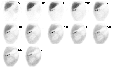
Sequential anterior abdominal images
View main image(hs) in a separate image viewer
Full history/Diagnosis is available below
Diagnosis: Intraperitoneal biliary leak
Full history:
12-year-old boy who is one month status post liver transplantation for cystic fibrosis. The
patient's biliary anastomosis is via a choledochocholedochostomy. The patient now presents with
mild jaundice (serum bilirubin 3.0 mg/dL) and increasing ascites. Paracentesis performed
the day before revealed an ascitic fluid bilirubin concentration of 27 mg/dL.
Approximately 5 liters of ascitic fluid was removed. The patient underwent
a T-tube cholangiogram on 5/5/98; this study revealed that the T-tube was
positioned out of the biliary tree, and the T-tube was subsequently
removed. Hepatobiliary scintigraphy was requested to confirm a biliary
leak.
Radiopharmaceutical:
1.2 mCi Tc-99m mebrofenin i.v.
Findings:
Following the intravenous administration of Tc-99m mebrofenin,
sequential anterior abdominal images were obtained through 60 minutes. There is
prompt, uniform accumulation of tracer by the liver. At 5 minutes
following administration of the radiopharmaceutical, a small amount of
activity is seen inferior to the tip of the right hepatic lobe. Over
the course of the remainder of the study, progressive accumulation of
activity is demonstrated throughout the peritoneal cavity, outlining
bowel loops. The scintigraphic findings are consistent with a biliary
leak. The exact site of the biliary leak is difficult to determine, since
there is lack of visualization of the intrahepatic biliary ducts and
common bile duct.
Note is also made of a linear region of activity with three more discrete
areas of tracer uptake just inferior to the right hepatic lobe; this
may represent accumulation of activity within the patient's T-tube tract.
Discussion:
The most common indication for hepatobiliary scintigraphy is clinical
suspicion of acute calculous or acalculous cholecystitis. In patients who have undergone biliary or hepatic
surgery, hepatobiliary scintigraphy may be used to evaluate for evidence
of a biloma or an intraperitoneal biliary leak. In these cases,
delayed imaging is often useful in making the diagnosis. In the case presented, evidence for an intraperitoneal biliary leak was
readily apparent early during imaging and no delayed images were
necessary.
Followup:
The patient underwent endoscopic retrograde cholangiopancreatography (ERCP) following
the hepatobiliary study. The ERCP reportedly revealed a leak in the
region of the distal bile duct. The leak appeared to
be contained and to be distal to the
choledochocholedochal anastomosis. A stent was subsequently placed
across the choledochocholedochal anastomosis.
ACR Codes and Keywords:
References and General Discussion of Hepatobiliary Scintigraphy (Anatomic field:Gasterointestinal System, Category:Organ specific)
Search for similar cases.
Edit this case
Add comments about this case
Return to the Teaching File home page.
Case number: hs013
Copyright by Wash U MO

