Case Author(s): Mike Roarke, M.D. and Henry D. Royal,M.D. , 3/8/96
3/8/96 . Rating: #D3, #Q4
Diagnosis: Congenital Common Bile Duct Obstruction.
Brief history:
2-month old boy with new onset
acholic stools and elevated GGT
Images:
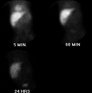
Anterior images of the abdomen.
View main image(hs) in a separate image viewer
View second image(us).
Sonogram showing gallbladder.
View third image(fl).
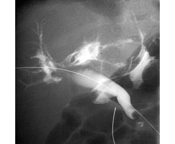
Transhepatic cholangiogram.
View fourth image(fl).
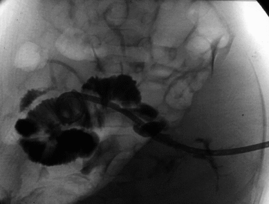
After placement of biliary catheter.
Full history/Diagnosis is available below
Diagnosis: Congenital Common Bile Duct Obstruction.
Full history:
2-month old boy with new onset
acholic stools and elevated liver enzymes. This
examination was requested to evaluate for evidence of
bile duct obstruction or atresia.
Radiopharmaceutical:
Tc-99m mebrofenin i.v.
Findings:
Hepatobiliary scintigraphy was
performed. At 5 minutes post injection, there is
prompt accumulation of tracer by the liver and some
residual blood pool activity within the heart. At 60
minutes, the heart blood pool activity is resolved and
the liver demonstrates no significant excretion into
the biliary tree. At 24 hours, there is still no biliary
tree visualization, no significant small bowel activity,
and there is some tracer within the urinary bladder
indicating vicarious excretion through the kidneys. A
sonographic image confirms the presence of a
gallbladder as a fluid filled structure adjacent to the
liver. Transhepatic cholangiogram demonstrates a
rounded mass-like structure producing obstruction to
contrast passage via the common bile duct. A follow-
up image demonstrates that a catheter was passed
beyond this obstruction and into the duodenum.
Discussion:
Following placement of the biliary
catheter, the obstructing lesion in the distal common
bile duct was no longer seen. It is felt that this
patient likely had a congenital web within the
common bile duct, which produced the obstruction to
bile flow. No evidence of a biliary calculus was found.
Differential Diagnosis List
In cases such
as this the major differential diagnosis includes
biliary atresia vs neonatal hepatitis. The prompt
hepatic uptake of tracer argues against neonatal
hepatitis, however. As this case demonstrates,
congenital bile duct obstruction can have an
appearance indistinguishable from that of biliary
atresia on hepatobiliary scintigraphy.
ACR Codes and Keywords:
References and General Discussion of Hepatobiliary Scintigraphy (Anatomic field:Gasterointestinal System, Category:Normal, Technique, Congenital Anomaly)
Search for similar cases.
Edit this case
Add comments about this case
Read comments about this case
Return to the Teaching File home page.
Case number: hs005
Copyright by Wash U MO