After viewing the image(s), the Full history/Diagnosis is available by using the link here or at the bottom of this page

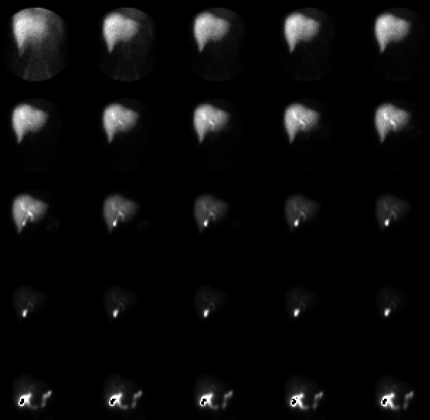
Sequential 2 minute images of the anterior abdomen. Images are scaled to the maximum pixel in each image; images in the last row are shown at greater intensity to show tracer in the bowel (resulting in grey-scale overflow elsewhere in the image)
View main image(hs) in a separate viewing box
View second image(hs). Images following administration of sincalide, displayed as sequential 2 minute frames for 30 minutes.
View third image(hs). A plot of activity in the region of the gallbladder after sincalide administration.
Full history/Diagnosis is also available
Return to the Teaching File home page.