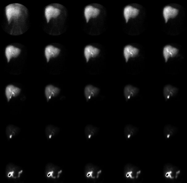

Sequential 2 minute images of the anterior abdomen. Images are scaled to the maximum pixel in each image; images in the last row are shown at greater intensity to show tracer in the bowel (resulting in grey-scale overflow elsewhere in the image)
View main image(hs) in a separate image viewer
View second image(hs). Images following administration of sincalide, displayed as sequential 2 minute frames for 30 minutes.
View third image(hs). A plot of activity in the region of the gallbladder after sincalide administration.
Full history/Diagnosis is available below
There was prompt contraction of the gallbladder following administration of 0.02 ug/k of sincalide by slow intravenous infusion. The calculated gallbladder ejection fraction was 50%. The sincalide infusion did not reproduce the patient's pain.
In this study, sincalide was administered to evaluate for chronic acalculus cholecystis. The normal response to sincalide argues against this disorder. However, the sensitivity and specificity of this "CCK HIDA" study is currently under debate in the literature.
References and General Discussion of Hepatobiliary Scintigraphy (Anatomic field:Gasterointestinal System, Category:Normal, Technique, Congenital Anomaly)
Return to the Teaching File home page.