Case Author(s): Samuel Wang, M.D. and Henry Royal, M.D. , 9/22/96 . Rating: #D2, #Q3
Diagnosis: Cavernous hemangioma
Brief history:
56-year old man with
prostate carcinoma and liver lesions identified
on CT study.
Images:
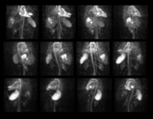
SPECT projection images.
View main image(hp) in a separate image viewer
View second image(hp).
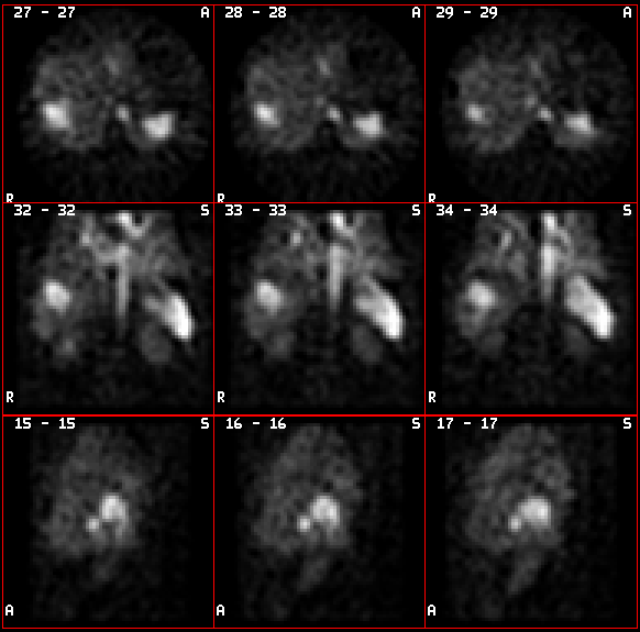
SPECT axial, coronal and sagittal images.
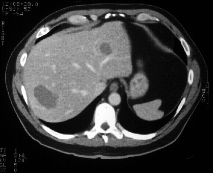
View third image(ct).
CT liver.
Full history/Diagnosis is available below
Diagnosis: Cavernous hemangioma
Full history:
This 56-year old man was
recently diagnosed with prostate carcinoma and
on a preoperative CT study was noted to have
at least two hypodense lesions in the liver,
which showed some peripheral nodular
enhancement suggestive of hemangiomas. The hepatic blood pool
study was obtained for confirmation.
Radiopharmaceutical:
Tc-99m in vitro
labeled red blood cells
Findings:
SPECT images were obtained
one hour post injection. These demonstrate
two foci of increased blood pool activity; the
smaller one in the left lateral lobe of the liver
and a second larger lobular one in the posterior
right lobe of the liver. These corresponded in
location to the hypodense lesions seen on the
CT study.
Discussion:
Hepatic blood pool scintigraphy has a very high predictive value for cavernous hemangiomas. Only a few false-positive cases have been reported. SPECT imaging improves contrast resolution and should be used when lesions are less than 3 cm in diameter or when there are multiple lesions.
ACR Codes and Keywords:
References and General Discussion of (Anatomic field:Gasterointestinal System, Category:Neoplasm, Neoplastic-like condition)
Search for similar cases.
Edit this case
Add comments about this case
Return to the Teaching File home page.
Case number: hp002
Copyright by Wash U MO