Case Author(s): Hamid Latifi,MD, Jerold Wallis, MD , 3/31/95 . Rating: #D2, #Q3
Diagnosis: Cavernous hemangioma
Brief history:
66 year old man with right upper abdominal pain
Images:

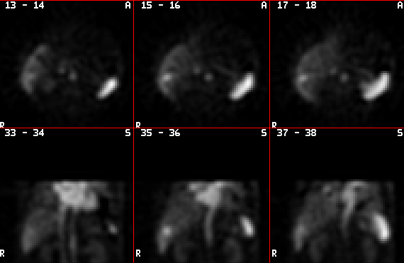
Transaxial(top row) and coronal(bottom row)
tomographic images obtained one hour after
administration of Tc-99m labeled red blood cells.
View main image(hp) in a separate image viewer
Full history/Diagnosis is available below
Diagnosis: Cavernous hemangioma
Full history:
66-year old man with elevated liver
function tests and right upper quadrant pain who had a
sonogram on 3-17-95 at an outside hospital. The sonogram
showed a hyperechoic lesion in the posterior segment of the
hepatic lobe, measuring approximately 3 cm. The current
examination was performed to confirm that this lesion
represents a benign cavernous hemangioma.
Findings:
Delayed tomographic images, demonstrate
increased blood pool activity in the lateral aspect of the
posterior segment of the right hepatic lobe, measuring
approximately 2.5-3 cm in diameter. This area of
scintigraphic abnormality corresponds to the sonographic
finding and is diagnostic of a cavernous hemangioma.
Discussion:
The sonographic findings in this case
were slightly atypical due to some areas of hypoechogenecity
within the central parts of this lesion. Therefore, hepatic
blood pool scintigraphy was recommended. On hepatic blood
pool imaging, the characteristic appearance of a cavernous
hemangioma is found. This appearance is extremely specific.
Major teaching point(s):
This case demonstrates the value of
hepatic blood pool scintigraphy in confirmation of hepatic
cavernous hemangioma. Tomographic images are especially
helpful in smaller lesions (less than 3-4 cm in diameter);
however, lesions less than 1.5 cm may not be visualized by
scintigraphic techniques.
Differential Diagnosis List
A single case of
hepatic angiosarcoma mimicking cavernous hemangioma has
been reported.
ACR Codes and Keywords:
References and General Discussion of (Anatomic field:Gasterointestinal System, Category:Neoplasm, Neoplastic-like condition)
Search for similar cases.
Edit this case
Add comments about this case
Read comments about this case
Return to the Teaching File home page.
Case number: hp001
Copyright by Wash U MO

