Case Author(s): Brock G. McDaniel, M.D. and Keith C. Fischer, M.D. , 11/17/02 . Rating: #D2, #Q3
Diagnosis: Diverticular bleed
Brief history:
71-year-old woman with maroon stools and decreasing hematocrit.
Images:

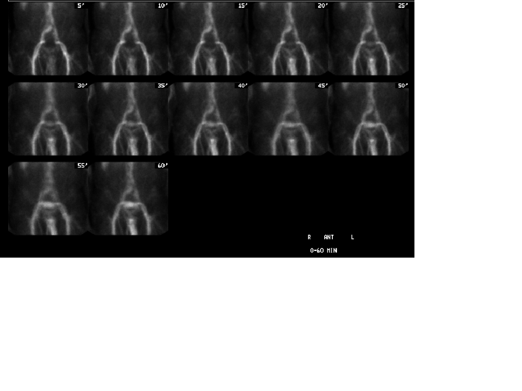
Anterior flow study
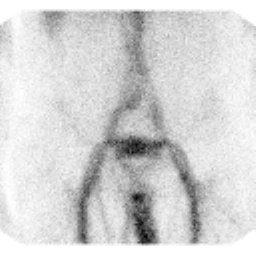
View main image(gi) in a separate image viewer
View second image(gi).
Anterior 0-60 minute (Animated GIF File)
View third image(gi).
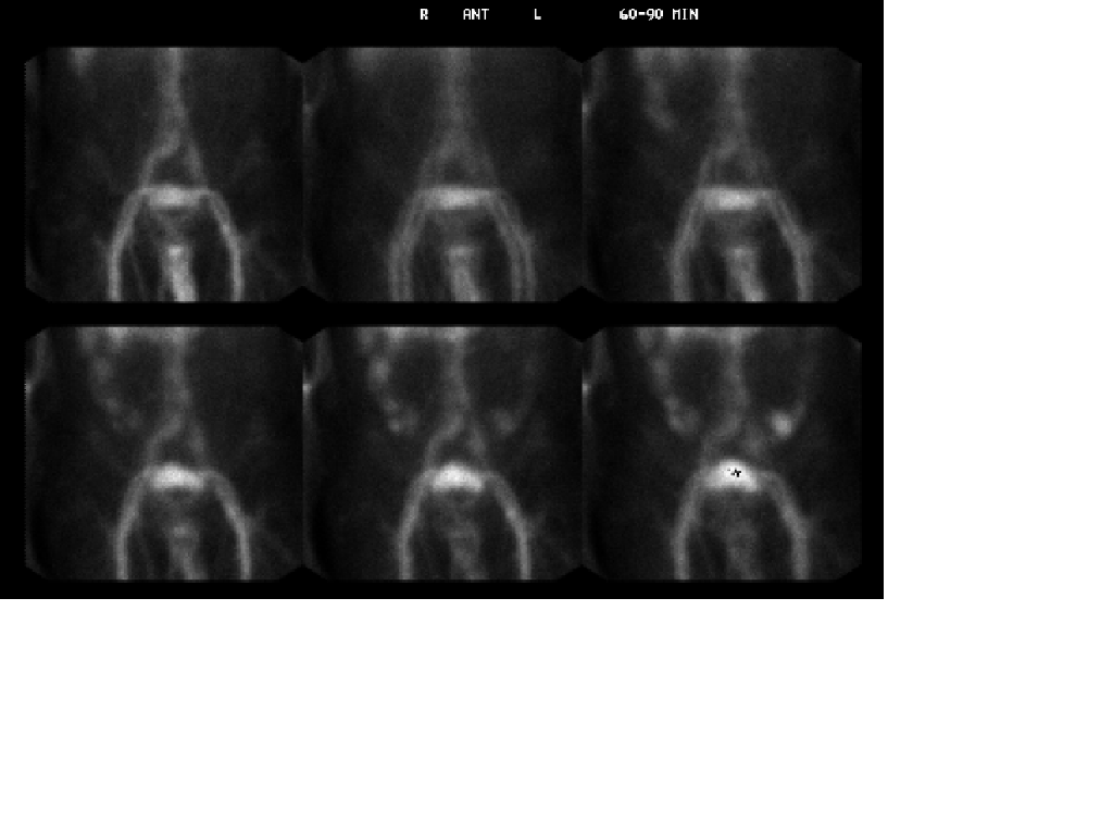
Anterior 60-90 minute (Animated GIF File)
View fourth image(an).
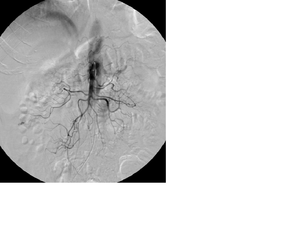
Selective SMA arteriogram
Full history/Diagnosis is available below
Diagnosis: Diverticular bleed
Full history:
71 year-old male with past history of diverticular disease status post left hemicolectomy for diverticular bleed, diabetes, and hypertension who was seen in the emergency room after having six mixed bloody and black stools. Subsequent tagged RBC study demonstrated active bleeding within the colon at the hepatic flexure. Vascular interventional radiology then demonstrated active bleeding from a branch of the right colic artery. Hemostasis was achieved with embolization utilizing three 2mm micro-coils. The following day the patient continued to have bright red blood per rectum, and therefor, the patient underwent a repeat tagged RBC bleeding study which again showed active bleeding at the hepatic flexure. The patient then was sent to surgery for a complete colectomy with an ileorectal anastomisis.
Radiopharmaceutical:
25.5 mCi Tc99m in vitro labeled red cells i.v.
Findings:
Flow images are normal. Early (0-60 minute) tagged RBC images are negative for active bleeding. Delayed (60-90 minute) images demonstrate a site of active bleeding located at the hepatic flexure with activity increasing in intensity over time, peristalsing distally and also moving dependently in a retrograde fashion to pool in the cecum. A subsequent selective right colic arteriogram demonstrates active extravasation consistent with a diverticular bleed. Hemostasis was achieved with coil embolization. However, the following day the patient returned with active bleeding which a repeat tagged RBC scan confirmed was originating from the hepatic flexure (not shown).
Discussion:
The rationale behind gastrointestinal bleeding studies is to determine if the patient is actively bleeding and if so to help guide the referring physicians in the next step of the patient’s management. Localization of the site of bleeding can help aid the interventional radiologist in determining the appropriate vessels for selective arteriography prior to embolization. Though the most often sited indication for performing a gastrointestinal bleeding study by referring physicians is bright red blood per rectum, orthostatic hypotension is clearly the most sensitive clinical indicator. Bright red blood per rectum is not a good indicator of acut bleeding because the time of colonic evacuation may not coincide with active bleeding.
Major teaching point(s):
It is always important to not rely only on the static planar images when trying to localize an active site of bleeding. If only static planar images were relied upon in this case, one could potentially mis- interpret the site of bleeding as originating from the cecum. Only after evaluating the dynamic cine images does it become apparent that the bleeding originates in the region of the hepatic flexure and reaches the cecum through dependent pooling.
ACR Codes and Keywords:
References and General Discussion of Gastrointestinal Bleeding Scintigraphy (Anatomic field:Gasterointestinal System, Category:Organ specific)
Search for similar cases.
Edit this case
Add comments about this case
Return to the Teaching File home page.
Case number: gi009
Copyright by Wash U MO