Case Author(s): Matt Jaksha, M.D. and Henry Royal, M.D. , . Rating: #D3, #Q4
Diagnosis: Gastric bleed
Brief history:
70 year old female with blood per rectum
Images:

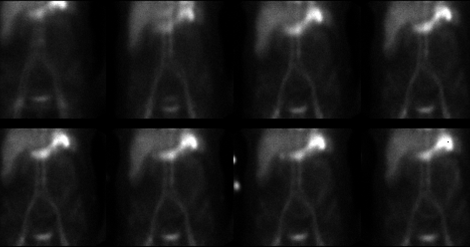
Sequential anterior images of the abdomen and pelvis through 50 minutes.
A cine file is available in AVI format.
View main image(gi) in a separate image viewer
View second image(gi).
Further anterior images though 90 minutes. A cine file is available in AVI format.
View third image(gi).
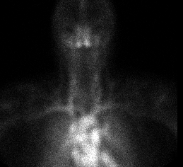
Anterior image of the neck
Full history/Diagnosis is available below
Diagnosis: Gastric bleed
Full history:
This is a 70 year old woman, two weeks status post myocardial infarction and LAD stent placement. She was anticoagulated, but the Coumadin was discontinued 4 days ago due to a drop in her hematocrit level. She presents now with blood per rectum and a decreasing hematocrit requiring blood transfusion.
Radiopharmaceutical:
20.4 mCi Tc-99m in vitro labeled red cells i.v.
Findings:
As early as 5 minutes into the study, an abnormal focus of activity is seen in the left upper quadrant. This subsequently increases in both size and intensity, and eventually defines the stomach. An image over the thyoid gland shows only blood pool activity in the neck, with no specific uptake in the thyroid gland.
Discussion:
Activity in the stomach on GI bleeding scintigraphy may represent free Tc-99m pertechnetate. In this case there were two clues that the stomach activity was not due to free pertechnetate. First, the initial activity appears to be pooling in the fundus of the stomach rather than accumulating within the wall of the stomach. Second, the activity within the stomach gradually increases; with free pertechnetate the activity should appear more rapidly (within 5-10 minutes after the injection). To confirm that free pertechnetate was not responsible for the findings, an image over the head and neck was obtained and no thyroid and salivary gland activity was seen.
Followup:
The patient underwent upper endoscopy, where a venous site of hemmorhage was identified in the gastric fundus.
Major teaching point(s):
Red blood cell scintigraphy is not indicated in patients with known upper GI bleeding as the only likely source for their bleeding. Occasionally a naso-gastric aspirate is not performed initially or is falsely negative.
Differential Diagnosis List
Free Tc-99m pertechnetate secretion in the wall of the stomach
ACR Codes and Keywords:
References and General Discussion of Gastrointestinal Bleeding Scintigraphy (Anatomic field:Gasterointestinal System, Category:Organ specific)
Search for similar cases.
Edit this case
Add comments about this case
Read comments about this case
Return to the Teaching File home page.
Case number: gi007
Copyright by Wash U MO