Case Author(s): Matt Jaksha, M.D. and Henry Royal, M.D. , . Rating: #D2, #Q3
Diagnosis: Portal hypertension with varices
Brief history:
64 year old male with coffee ground emesis.
Images:

Multiple images from gastrointestinal bleeding scintigraphy
View main image(gi) in a separate image viewer
Full history/Diagnosis is available below
Diagnosis: Portal hypertension with varices
Full history:
This is a 64 year old male with a history of nausea, coffee ground emesis and melena. EGD revealed an esophageal ulcer which was not actively bleeding. Scintigraphy was requested to evaluate any lower gastrointestinal bleed.
The pertinent past medical history included alcoholic hepatitis with cirrhosis and esophageal varices.
Radiopharmaceutical:
21.0 mCi Tc-99m in vitro labeled red cells i.v.
Findings:
No abnormal foci are seen to indicate gastrointestinal bleeding. The central acculmulation of activity within the abdomen suggests ascites, and there are numerous ectatic vessels consistent with mesenteric varices.
Discussion:
This patient's presentation and history strongly suggest bleeding from esophageal varices. Variceal bleeding can be life-threatening. There is often no obvious precipitating event. Fifty percent will resolve with no therapy, but the rate of recurrent bleeding is high.
Followup:
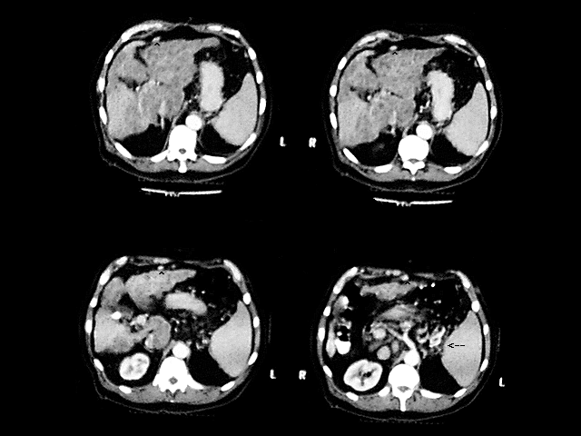
Comparison with prior CT of the abdomen confirmed cirrhotic changes. Besides ascites, multiple collateral vessels are seen. These include a recanalized periumbilical vein and splenic hilar collaterals. The spleen is enlarged and the liver is nodular, with hypertrophy of the caudate lobe.
View followup image(ct).
Abdominal CT images 1 year prior to GI bleeding scintigraphy
Major teaching point(s):
GI bleeding studies occasionally demonstrate findings which do not indicate bleeding but are significant (e.g. cases of undiagnosed colon or renal cell carcinoma).
ACR Codes and Keywords:
References and General Discussion of Gastrointestinal Bleeding Scintigraphy (Anatomic field:Gasterointestinal System, Category:Organ specific)
Search for similar cases.
Edit this case
Add comments about this case
Read comments about this case
Return to the Teaching File home page.
Case number: gi006
Copyright by Wash U MO