Case Author(s): J. Philip Moyers, MD, and Barry A. Siegel, MD , 1/17/96 . Rating: #D3, #Q3
Diagnosis: Occlusion of the superior vena cava
Brief history:
The patient presented with
hematochezia on the evening before this
study. The patient now has persistent melena.
Please evaluate for source of
bleeding.
Images:

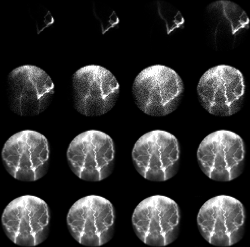
Anterior summed images from GI bleeding study
View main image(gi) in a separate image viewer
Full history/Diagnosis is available below
Diagnosis: Occlusion of the superior vena cava
Full history:
This patient has a history of
lymphoma and known superior vena caval obstruction
secondary to thrombosis caused by indwelling central venous access
catheters. The patient does not currently have
superior vena cava (SVC) syndrome.
Radiopharmaceutical:
20.6 mCi Tc-99m in
vitro labeled red blood cells
Findings:
The anterior radionuclide
angiogram demonstrates abnormal activity over the
left flank extending into the left abdominal region and
finally to the left pelvis, then traversing in a more
caudad direction prior to visualization of the aorta
and iliac artery. This study was done after a left arm
injection of the radiopharmaceutical. Delayed images
demonstrate multiple irregular linear collections of
activity along the abdominal wall consistent with
dilated anterior abdominal wall collateral venous
structures.. These findings are consistent with
superior vena caval obstruction with abdominal wall
collateral vasculature. No abnormal focus of labeled red blood cell
extravasation
is demonstrated to suggest gastrointestinal bleeding,
however.
Discussion:
Superior vena cava syndrome, which is
caused by obstruction of the SVC with development of
collateral pathways, is characterized by head and neck edema
(70%), enlarged cutaneous venous collaterals, syncope,
headache, dizziness, proptosis, excessive tearing, dyspnea,
chest pain, cyanosis, and hematemesis (11%). The causes of
superior vena cava obstruction may be divided into malignant
(85%) and benign (15%) etiologies. Bronchogenic carcinoma
and lymphoma make up the majority of malignant causes.
Benign diseases causing SVC obstruction include
granulomatous mediastinitis (tuberculosis, histoplasmosis, sarcoidosis,
etc.), substernal goiter, aortic aneurysm, constrictive
pericarditis, and foreign bodies (central venous catheters/pacer
wires).
Radiographic findings include superior mediastinal widening,
dilated cervical and superficial veins, SVC thrombus, and
encasement or compression or occlusion of the SVC. The
common collaterals pathways include the esophageal venous
plexus (downhill varices), azygous and hemiazygous veins,
accessory hemiazygous and superior intercostal veins (aortic
nipple), lateral thoracic veins, parumbilical veins, and vertebral
veins.
In SVC obstruction, Tc-99m sulfur colloid scintigraphy (not
performed here) often displays a characteristic pattern. The
intravenously injected radiopharmaceutical passes through
collateral thoracic veins to abdominal wall collaterals and
thence to the paraumbilical veins, if patent. The paraumbilical veins
usually drain into a branch of the left portal vein supplying the
quadrate
lobe of the liver. Under these circumstances, liver-spleen scintigraphy shows a
focal area of increased Tc-99m sulfur colloid uptake in this region of the liver.
Followup:
The patient was taken to the
angiography suite where recannulization of an
occluded right-sided subclavian vein was performed.
A superior vena cavogram at that point demonstrated
extensive SVC thrombus extending into the right atrium.
Because of this, no superior vena caval stent was
placed.
ACR Codes and Keywords:
References and General Discussion of Gastrointestinal Bleeding Scintigraphy (Anatomic field:Vascular and Lymphatic Systems, Category:Organ specific)
Search for similar cases.
Edit this case
Add comments about this case
Read comments about this case
Return to the Teaching File home page.
Case number: gi003
Copyright by Wash U MO

