Case Author(s): Sarah Reimer, MD , Keith Fischer MD, and Farrokh Dehdashti, MD , 03/19/99 . Rating: #D3, #Q4
Diagnosis: Non-Hodgkin's Lymphoma
Brief history:
S/p bone marrow transplant for NHL, now with fever and malaise.
Images:
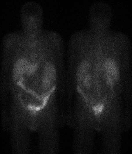
Anterior and posterior whole body gallium images obtained at 72 hours
View main image(ga) in a separate image viewer
View second image(pt).
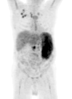
Projection image from FDG PET scan
View third image(pt).
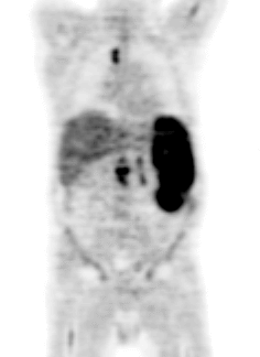
Coronal image from FDG PET scan
Full history/Diagnosis is available below
Diagnosis: Non-Hodgkin's Lymphoma
Full history:
This is a patient with a high-grade NHL, s/p bone marrow transplantation
4 months previously. He presented with a palpable spleen, pancytopenia,
dyspnea, and fever. The patient had had an MRI with gadolinium one week
prior to the Gallium scan. The MRI showed findings most consistent with
multiple peripheral splenic infarcts, increasing splenomegaly, and no
change in multiple small retroperitoneal lymph nodes.
Radiopharmaceutical:
Gallium-67 citrate i.v.
F-18 fluorodeoxyglucose (FDG) i.v.
Findings:
The Gallium scan demonstrated splenomegaly without focal increased
activity to suggest tumor and no evidence of residual nodal disease.
The FDG-PET scan demonstrated splenomegaly with intensely increased
activity. Multiple right axillary, mediastinal, mesenteric, celiac,
and retroperitoneal lymph nodes were also intensely FDG-avid. The liver
had heterogeneous uptake and there was diffusely increased activity in
the bone marrow.
Major teaching point(s):
FDG-PET can be used to more reliably detect recurrent/residual lymphoma.
ACR Codes and Keywords:
- General ACR code: 98
- Vascular and Lymphatic Systems:
9.8343 "Non-Hodgkin lymphoma"
References and General Discussion of Gallium Scintigraphy (Anatomic field:Vascular and Lymphatic Systems, Category:Misc)
Search for similar cases.
Edit this case
Add comments about this case
Read comments about this case
Return to the Teaching File home page.
Case number: ga006
Copyright by Wash U MO