Case Author(s): Dennis Hsueh, M.D. and Tom R. Miller, M.D., Ph.D. , 10/8/03 . Rating: #D2, #Q3
Diagnosis: Leakage of cerebral spinal fluid from ventriculo-peritoneal shunt
Brief history:
37 year old woman with worsening headaches and developing mass in the left neck.
Images:
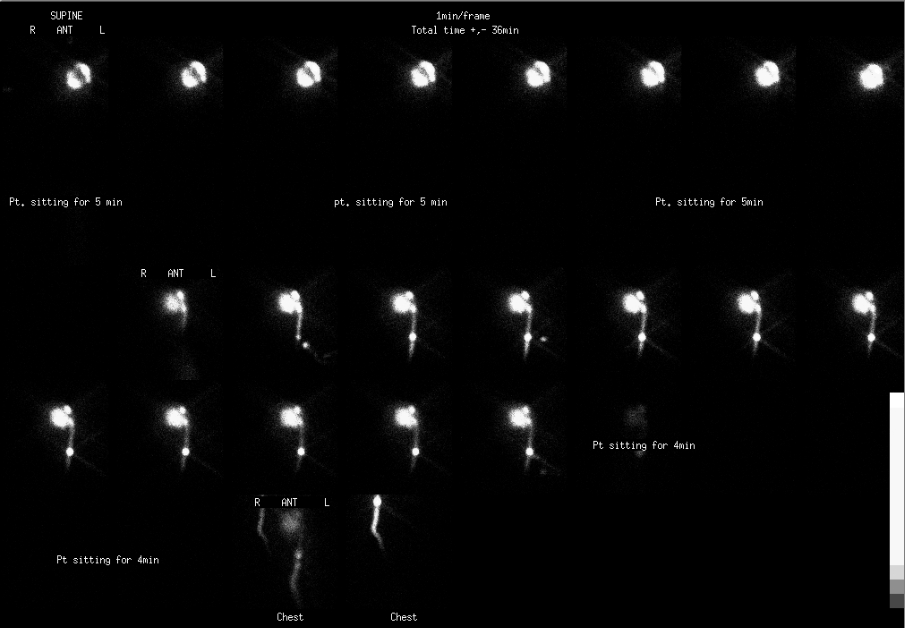
Anterior images. Top row images (obtained with patient supine); middle two row images (obtained after patient sitting for 5 minutes); bottom two images (obtained after patient sitting for another 4 mintues)
View main image(cs) in a separate image viewer
Full history/Diagnosis is available below
Diagnosis: Leakage of cerebral spinal fluid from ventriculo-peritoneal shunt
Full history:
37 year old woman with a history of Arnold-Chiari malformation and arachnoid cyst treated with posterior fossa decompression and cerebrospinal fluid (CSF) shunt placement 24 years ago. She has had multiple revisions of her CSF shunts and now presents with worsening headaches and a developing erythematous swelling in her left neck. There was clinical concern for malfunction of her CSF shunt.
Radiopharmaceutical:
Tc-99m DTPA injected into CSF resevoir
Findings:
Initial images were obtained following injection of the reservoir following manual compression of the distal limb of the CSF diversionary shunt. The images demonstrate reflux of radiopharmaceutical into the ventricular system indicating patency of the proximal limb.
Flow into the shunt improves after the patient sits for 5 minutes. The subsequent images demonstrate flow into the distal limb in the head, neck and upper chest. A more intense focal collection in the shunt localizes at the site of the patient’s focal neck swelling.
Because images confirmed that a CSF leak site was present in the neck, images to evaluate the intra-peritoneal segment of the shunt were not performed.
Discussion:
Malfunction of the CSF shunt most commonly occurs from obstruction. The presenting symptoms may be nonspecific: nausea and vomiting, headache, and irritability. Causes of obstruction are variable ranging from fibrin debris, blood clot, and ingrowth by choroid or glial elements. Loculation in the distal segment by abdominal adhesions can lead to a CSF pseudocyst. The patency of shunts can be evaluated with scintigraphy. See teaching file case CS001 for discussion of a normal examination.
Although CSF leakage can occur following trauma, typical sites are from the paranasal or mastoid sinuses. Leakage of CSF from a shunt, a more rare occurrence, can be due to catheter disconnecting from the resevoir or catheter fracture. Leaks can be intermittent and can occur with changes in patient position. Findings with a CSF leak are “extravasation” or collections of radiotracer in a abnormal location outside the cerebrospinal space.
In this case, the patient’s abnormal neck swelling was due to leakage of CSF from a disrupted shunt.
Followup:
Although this examination demonstrated patency of the shunt beyond the leak site, because of the risk of infection and obstruction, the patient was brought in for surgical revision of the shunt. A partial fracture in the catheter was confirmed at surgery.
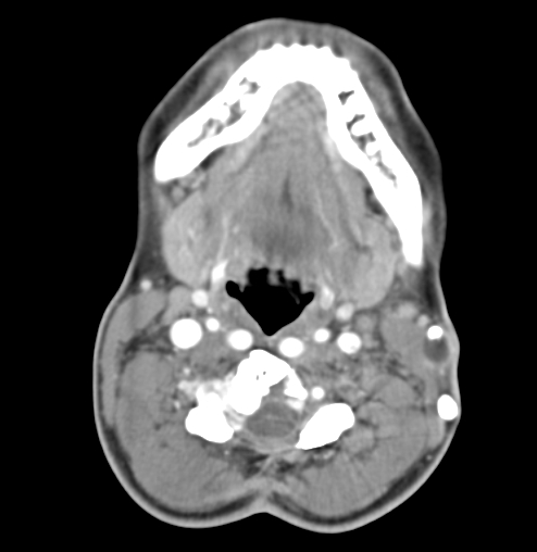
View followup image(ct).
This axial CT examination was obtained the same day as the CSF shunt scintigraphy examination. This select axial post contrast CT of the neck was obtained in the region of the left neck swelling. Note fluid density collection posterior and adjacent to left shunt tubing, superficial to the left sternocleidomastoid muscle. Incidentally noted is an older, nonfunctioning shunt posterior to this malfunctioned shunt. No cervical adenopathy was appreciated.
ACR Codes and Keywords:
References and General Discussion of CSF Shunt Scintigraphy (Anatomic field:Spine and Contents, Category:Misc)
Search for similar cases.
Edit this case
Add comments about this case
Return to the Teaching File home page.
Case number: cs005
Copyright by Wash U MO