Case Author(s): Brock G. McDaniel, M.D. and Tom R. Miller, M.D., Ph.D. , 12/09/02 . Rating: #D3, #Q4
Diagnosis: Obstructed ventriculo-atrial CSF shunt
Brief history:
23 year old male with Chiari 2 malformation and spina bifida who presents with worsening headache.
Images:
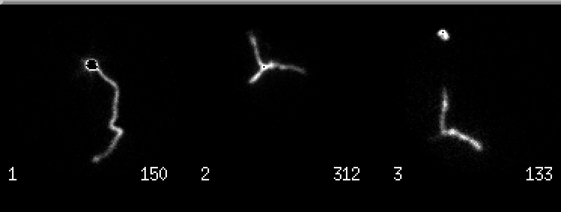
Anterior images: immediate head/neck, 5 minute chest, and 1 hour head/neck/chest
View main image(cs) in a separate image viewer
View second image(xr).
PA chest radiograph
View third image(ct).
Non-contrast head CT
View fourth image(an).
Left upper extremity venogram
Full history/Diagnosis is available below
Diagnosis: Obstructed ventriculo-atrial CSF shunt
Full history:
The patient is a 23 year old man with congenital hydrocephalus and a ventricuol-atrial shunt who presented with worsening headache and mental status changes. He has had multiple shunt placements and revisions in the past. During one of his previous episodes of obstruction he had similar symptoms including worsening headache.
Radiopharmaceutical:
0.47 mCi Tc-99m DTPA injected into the reservoir of the shunt system by the neurosurgery service.
Findings:
On both the immediate and 5-minute delayed images, there is prompt activity within the shunt system from the level of the reservoir to the region of the sternal notch. A delayed 60-minute image demonstrates bifurcation of the activity with no significant clearance into the right atrium. The plain film shows the shunt to be intact with the tip positioned at approximately the superior venacaval/brachiocephalic vein junction. The head CT demonstrates stable, mild to moderate hydrocephalus and a left ventriculostomy catheter. The left upper extremity venogram demonstrates occlusion of the left subclavian and brachiocephalic veins. Numerous collaterals at the base of the left neck are also seen.
Discussion:
The patency of a ventriculo-peritoneal or ventriculo-atrial shunt can be assessed by injecting a small amount (typically about 0.2ml) of radiopharmaceutical into the shunt reservoir. The normal transit time is approximately 10-20 minutes. Typically the patient is supine for the injection. To increase hydrostatic pressure, the patient can be imaged upright. When injecting the reservoir, it is important not to withdrawal CSF, thereby reducing the pressure in the system; doing so could lead to an erroneous conclusion of a partial obstruction.
Followup:
The patient was subsequently taken to the operating room where the left ventriculo-atrial shunt was removed and a new right ventriculo-atrial shunt was placed. By post-operative day 2 the patient had returned to his baseline mental status and reported resolution of his headache.
ACR Codes and Keywords:
References and General Discussion of CSF Shunt Scintigraphy (Anatomic field:Skull and Contents, Category:Other generalized systemic disorder)
Search for similar cases.
Edit this case
Add comments about this case
Return to the Teaching File home page.
Case number: cs004
Copyright by Wash U MO