Case Author(s): Anton J. Johnson, M.D., Ph.D., and Jerold W. Wallis, M.D. , 4/15/97 . Rating: #D3, #Q5
Diagnosis: Infiltrated injection
Brief history:
36 year old woman with a ventriculoperitoneal shunt presents with
headache, diplopia, and seizures.
Images:

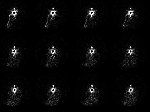
Initial 30-second anterior images obtained with the camera over the
patient's head and neck.
View main image(cs) in a separate image viewer
View second image(cs).
Continuation of image set one with additional images of the abdomen.
View third image(cs).
Same images as the first set only displayed at higher intensity.
Full history/Diagnosis is available below
Diagnosis: Infiltrated injection
Full history:
36 year old woman with a history of hydrocephalus and
ventriculoperitoneal shunt placement presents with headache,
diplopia, and seizures. The CSF shunt study was requested to evaluate
for evidence of shunt malfunction or obstruction.
Radiopharmaceutical:
0.20 mCi Tc-99m DTPA (infiltrated dose); and a second dose of 0.88 mCi
Tc-99m DTPA injected into the shunt reservoir.
Findings:
The initial images following the first injection show no movement of
radiopharmaceutical down the ventriculoperitoneal shunt tubing. The
activity appears to remain in the reservoir. An image of the abdomen
reveals renal excretion of activity, a finding consistent with
significant systemic exposure to the dose.
Displaying the initial images at higher intensity allows visualization
of tiny lymphatic channels draining activity away from an infiltrated
dose. A progressive increase in soft tissue activity is also visible;
normally, little or no soft tissue activity is visible if the dose has
been properly injected into the reservoir.
Followup:
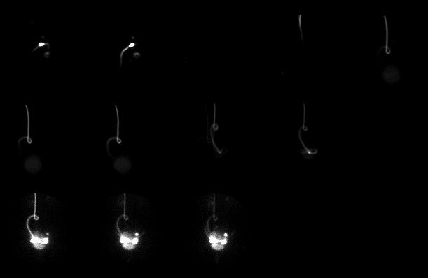
The patient underwent a second injection, this time into the shunt
reservoir. The initial images (shown below)
show activity in the reservoir,
with some reflux of activity into the lateral ventricle.
Subsequent images demonstrate a patent ventriculoperitoneal shunt with
mildly delayed transit (since the tracer required nearly an hour to
reach the end of the shunt tubing).
View followup image(cs).
Sequential 5 min images after re-injection.
Major teaching point(s):
If there is no movement of activity on a CSF shunt study, be sure to
exclude the possibility of an infiltrated injection before diagnosing
shunt obstruction.
An alternate method of performing this examination involves intentional
reflux into the ventricular system. At this institution, we generally do
not try to reflux into the ventricles. Instead, we introduce
the tracer into the shunt system in a very small volume, and observe
for progressive flow. If there is flow, it implies that both the
proximal and distal limbs must be patent.
ACR Codes and Keywords:
References and General Discussion of CSF Shunt Scintigraphy (Anatomic field:Skull and Contents, Category:Normal, Technique, Congenital Anomaly)
Search for similar cases.
Edit this case
Add comments about this case
Read comments about this case
Return to the Teaching File home page.
Case number: cs003
Copyright by Wash U MO