Case Author(s): Michael Roarke, M.D. and Barry A. Siegel, M.D. , 3/9/96 . Rating: #D3, #Q4
Diagnosis: Obstructed Ventriculoperitoneal Shunt
Brief history:
3-month old girl with
hydrocephalus and loculated fluid collection in the
abdomen on ultrasonography.
Images:
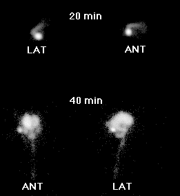
Anterior and lateral images of the head.
View main image(cs) in a separate image viewer
Full history/Diagnosis is available below
Diagnosis: Obstructed Ventriculoperitoneal Shunt
Full history:
3-month old girl with history of
jejunal atresia status post surgical repair and
subsequent development of hydrocephalus. A
ventriculoperitoneal shunt was placed on 12-5-95,
complicated by pseudocyst formation at the distal tip
of the peritoneal limb secondary to adhesions.
Radiopharmaceutical:
Tc-99m DTPA
Findings:
At 20 minutes following injection of
tracer into the shunt reservoir, activity is seen in the
proximal limb tubing only. At 40 minutes, activity is
seen in the lateral ventricles, and third and fourth
ventricles, as well as extending down the spinal canal.
Still, no activity is seen in the distal shunt limb.
Sonographic examination confirms the presence of a
loculated fluid collection within the abdomen at the
site of the distal limb tip.
Discussion:
Patency of the distal limb of a
ventriculoperitoneal shunt catheter is indicated by
free peritoneal spill of activity. The absence of distal
limb tracer activity at 40 minutes in this examination,
despite pumping the reservoir valve, indicates distal
limb obstruction. The patient subsequently
underwent conversion to a ventriculoatrial shunt.
ACR Codes and Keywords:
References and General Discussion of CSF Shunt Scintigraphy (Anatomic field:Skull and Contents, Category:Normal, Technique, Congenital Anomaly)
Search for similar cases.
Edit this case
Add comments about this case
Return to the Teaching File home page.
Case number: cs002
Copyright by Wash U MO