Case Author(s): Thomas H. Vreeland, MD / J. Wallis , 07/17/94 . Rating: #D2, #Q3
Diagnosis: Normal CSF shunt scintigraphy
Brief history:
The patient is being evaluated for possible
shunt malfuntion.
Images:
View main image(cs) in a separate image viewer
Full history/Diagnosis is available below
Diagnosis: Normal CSF shunt scintigraphy
Full history:
This patient is a four year old boy with a
history of resection of a posterior fossa tumor and place-
ment of a ventriculo-peritoneal shunt catheter system two
years ago. The patient now presents with nausea, vomiting,
headache and lethargy. The patient is being evaluated
for possible shunt malfunction.
Radiopharmaceutical:
0.5 mCi Tc-99m DTPA in less than 0.2 ml.
Note: Tc-99m DTPA is not specifically approved for intrathecal
administration, and accordingly meets the phamacopoeial bacterial
endotoxin standard for an intravenously administered drug. Because the
CNS is more sensitive to endotoxins, only < 10% of the volume of a Tc-
99m DTPA vial should be injected for this study whenever the
radiopharmaceutical is to be refluxed into the ventricular system. In this case, there was no intention to reflux tracer into the ventricles.
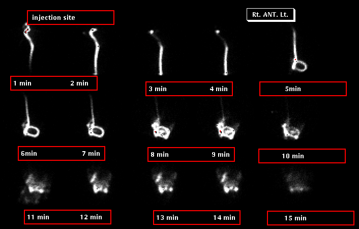
Findings:
There is normal progression of tracer down the distal shunt
catheter system, with free spillage of tracer activity into
the peritoneal cavity by 15 minutes. There is no reflux
into the ventricles.
Discussion:
(1) At our institution, the Neurosurgeon will inject the
radiopharmaceutical directly into the shunt catheter
reservoir system. The total volume of tracer
is 0.2 to 0.4 ml., injected with a tuberculin syringe.
A small volume is
used to avoid disturbing flow in the system. The patient
is usually imaged in
the supine position. Dynamic one minute images are acquired,
and additional analog images are acquired.
(2) The normal transit time is approximately 10-20 minutes.
A transit time of more than 30 minutes is
abnormal. Althought the proximal limb is not evaluated
directly, if there is normal flow through the system then
the proximal limb must be patent.
(3) Removal of fluid (e.g. for culture) is avoided, as it
may decrease the pressure below that needed to open the
shunt valve and may artifactually produce an abnormal
test result.
(4) If no flow is seen, placing the patient in an erect
position will slightly increase the
forward pressure and encourage CSF flow.
ACR Codes and Keywords:
References and General Discussion of CSF Shunt Scintigraphy (Anatomic field:Skull and Contents, Category:Organ specific)
Search for similar cases.
Edit this case
Add comments about this case
Read comments about this case
Return to the Teaching File home page.
Case number: cs001
Copyright by Wash U MO