Case Author(s): Rusty Roberts M.D. and Keith Fischer M.D , 11/26/04 . Rating: #D3, #Q3
Diagnosis: Marfan's Syndrome with dural ectasia
Brief history:
47 year old woman has severe headaches, and the concern is for leak with cerebral spinal fluid hypotension as the etiology for the headaches
Images:
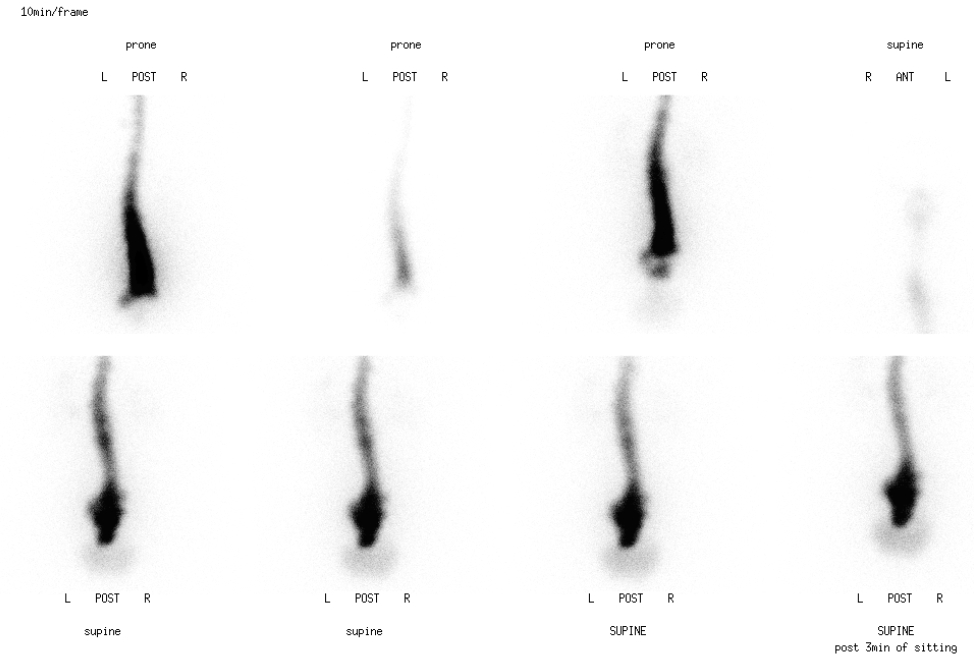
Immediate post subarachnoid injection of the radiopharmaceutical--Planar images of the spine
View main image(cl) in a separate image viewer
View second image(cl).
Lateral and PA planar scintigrams
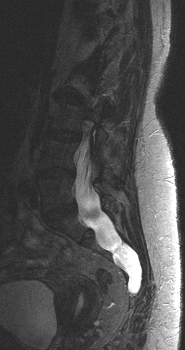
View third image(mr).
Axial T2 weighted MR images. View MR cine in AVI format.
View fourth image(mr).
Sagittal T2 weighted MR images. View MR cine in AVI format.
Full history/Diagnosis is available below
Diagnosis: Marfan's Syndrome with dural ectasia
Full history:
47 year old woman with Marfan's syndrome has evidence of dural ectasia with neural cysts on MRI. She has severe headaches and the concern is for leak with CFS hypotension as the etiology.
Radiopharmaceutical:
0.488 mCi In-111 DTPA by lumbar subarachnoid injection at L3-L4
Findings:
The radiopharmaceutical was injected at the L3-L4 lumbar subarachnoid level by staff of the Section of Neuroradiology. The patient was brought immediately to the imaging suite in the prone position and three 10-minute images obtained.
These images demonstrate a widened subarachnoid space in the lumbar and sacral region. There is evidence of bladder activity during the second 10-minute image. The kidneys are clearly visible on the third 10-minute image. The patient was then placed in the supine position and imaging continued. The pattern of activity did not change. There is still some widening of the subarachnoid space in the lower lumbar spine and sacrum. Bladder activity continued to be seen. The patient sat up for 3 minutes, and imaging following this showed no change in the pattern of activity. The patient then walked to the bathroom, urinated, and walked back.
Additional images showed no change in the pattern of subarachnoid activity. Imaging over the head confirmed the subarachnoid location of activity. Lateral imaging in the lower lumbar spine and sacrum showed no activity posterior or anterior to the subarachnoid space.
MRI images delineate the extent of the dural ectasia seen on the scintigraphic images. The apparent decrease signal within the CSF on the axial images is an artifact from the file compression.
Discussion:
Marfan's Syndrome is an inherited (autosomal dominant) disorder, caused by a defective gene involved with the production of fibrillin. Fibrillin makes up part of connective tissue in the body, such as the blood vessels, eye lenses, ligaments, and dura. Dural ectasia is also seen with perineural cysts and meningeal cysts. Dural ectasia may be seen in 41%, but when it occurs, 99% are in the lower spine. It is postulated to be caused by stretching of the dura over time. Pain management is therapy, also Diamox (acetazolamide) has been used. (Ahn, N. Johns Hopkins 2001)
The symptoms of dural ectasia vary. They include low back and abdominal pain, headaches and leg pain. Perineal pain and numbness can also occur because of the sacral nerve roots involvement.
Spontaneous intracranial hypotension from a CSF leak in a patient with Marfan’s syndrome. Journal of Neurology, Neurosurgery & Psychiatry. 59(5):516-9. 1995 Nov.
Dural ectasia in the Marfan syndrome: MR and CT findings and criteria. Genet Med. 2000 May-Jun;2(3):173-9.
Followup:
The rapid appearance of bladder activity on the immediate post injection images, and the lumbar dural ectasia led physicians to suspect a lumbar CSF leak. The patient was debilitatingly symptomatic and wished to try a blood patch. This was performed the day following the nuclear scintigraphy. Within one day, the patients symptoms, nausea and headache, had improved. At the two month follow up appointment, her symptoms had almost completely resolved.
Differential Diagnosis List
cerebral spinal fluid leak with dural ectasia, meningeal cysts, or perineural (Tarlov) cysts
ACR Codes and Keywords:
References and General Discussion of Cerebral Spinal Fluid Leak (Anatomic field:Spine and Contents, Category:Other generalized systemic disorder)
Search for similar cases.
Edit this case
Add comments about this case
Return to the Teaching File home page.
Case number: cl004
Copyright by Wash U MO