Case Author(s): Mark Fister, M.D., and Tom R. Miller, M.D., Ph.D. , 9/21/2000 . Rating: #D2, #Q4
Diagnosis: CSF leak
Brief history:
Sixty year-old woman with recurrent episodes of meningitis presents with persistent rhinorrhea.
Images:
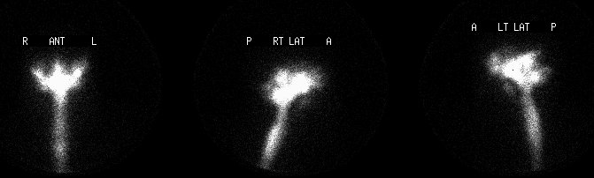
Four-hour delayed In-111 DTPA images of the head.
View main image(cl) in a separate image viewer
View second image(cl).
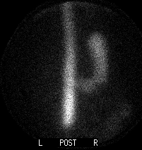
Four-hour delayed In-111 DTPA images of the abdomen.
View third image(cl).
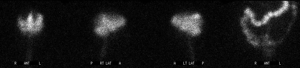
24-hour delayed images.
View fourth image(cl).
48-hour delayed images
Full history/Diagnosis is available below
Diagnosis: CSF leak
Full history:
Sixty year-old woman with recurrent episodes of meningitis presents with persistent rhinorrhea.
Radiopharmaceutical:
In-111 DTPA, intrathecal
Findings:
The 4-hour In-111 DTPA radionuclide cisternogram images demonstrate stomach and posterior nasal cavity/nasopharyngeal activity. Four-hour nasal pledget activity (normalized to blood activity) was markedly elevated on both sides, with greater activity on the left, suggesting a leak at that site. Twenty-four-hour delayed images demonstrate greater activity within the bowel and less prominent activity in the nasopharynx. Also noted is delayed transit of the tracer over the cerebral convexities. These findings persist on the 48-hour images, although the nasopharyngeal activity is barely perceptible at this time.
Discussion:
Cases of CSF rhinorrhea can be classified according to their cause. Although the vast majority are the result of direct head trauma, a substantial minority occur as a complication of surgery, and almost 5% are spontaneous. Approximately three-quarters of traumatic CSF leaks spontaneously resolve within 1 week, and nearly all within 6 months. Conversely, spontaneous leaks characteristically persist intermittently for years. The most serious complication is pyogenic meningitis which tends to occur in a higher percentage of postraumatic fistulas, although it also complicates a significant proportion of spontaneous leaks. Consequently, early surgical repair is appropriate, whatever the etiology.
There are various methods of presurgical imaging. Because the majority of CSF leaks produce only a small volume of fluid and are only intermittently active, radionuclide cisternography is the ideal imaging method for detecting and localizing a CSF leak. It is typically performed with the placement of pledgets (e.g., nasal pledgets in the case of CSF rhinorrhea) and complemented by the more precise localizing ability of directed, thin section CT. Cotton pledgets are labeled according to site, their activity counted separately in well counters, and weighed, with radioactivity normalized to gram of absorbed fluid and expressed as a ratio to plasma (because some of the radiopharmaceutical is absorbed at the arachnoid granulations and thereby becomes systemic). The pledget with the greatest activity is presumed to be the site closest to the CSF leak, and a pledget/plasma ratio of > 1.5 is interpreted as evidence of a CSF leak (typical ratios are much higher). Lateral images of the skull can directly identify leaks in some circumstances, and images of the abdomen (for swallowed activity) can be extremely useful, as in this case.
This case also demonstrates impaired ascent of the radiotracer over the cerebral convexities, presumably due to post-inflammatory changes in this patient with recurrent meningitis. The resulting altered CSF flow dynamics may have contributed to a spontaneous CSF leak, as a means of decompression. (the chicken or the egg?)
Reference: Lawrence SK, Sandler MP, Partain CL, and James AE, “Cerebrospinal Fluid Imaging” pp. 1163-1176 in Diagnostic Nuclear Medicine, 3rd ed. Sandler, et al. Williams and Wilkins, Baltimore, 1996.
Followup:
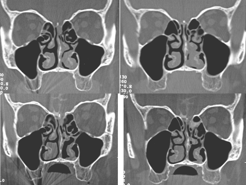
A thin-section CT cisternogram confirmed the presence of a 4 mm defect in the mid-left cribriform plate which was subsequently repaired.
View followup image(ct).
Coronal CT cisternogram images, precontrast above postcontrast images.
Major teaching point(s):
Radionuclide cisternography is an ideal imaging method for detecting and localizing a CSF leak, particularly when complemented by directed, thin-section CT.
ACR Codes and Keywords:
References and General Discussion of Cerebral Spinal Fluid Leak (Anatomic field:Skull and Contents, Category:Effect of Trauma)
Search for similar cases.
Edit this case
Add comments about this case
Return to the Teaching File home page.
Case number: cl003
Copyright by Wash U MO