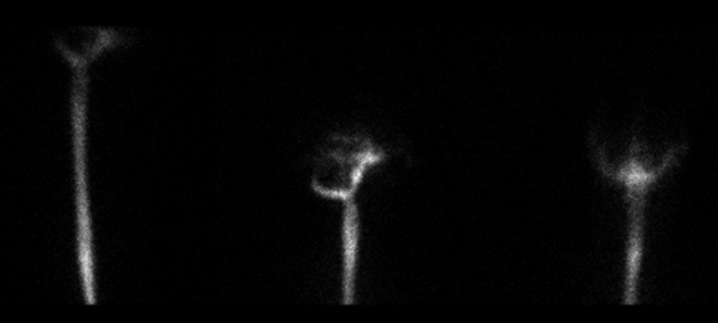
Images obtined 1 hour after the injection of the radiopharmaceutical into the lumbar subarachnoid space.
View main image(cl) in a separate image viewer
View second image(cl). Same 1 hr. image with increased intensity.
View third image(cl). 3 hr. delayed images over the abdomen.
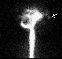
View fourth image(cl). 1 hr. lateral view over head with pointer.
Full history/Diagnosis is available below
Images obtained 1 hour after the injection of the radiopharmaceutical into the lumbar subarachnoid space reveals symmetric ascent of tracer in the cerebral subarachoid space. There is no ventricular reflux. A small amount of activity is seen anteriorly in the superior aspect of the nasopharynx (see 4th image.)
Anterior and posterior images over the abdomen were obtained approximately 3 hrs. after the injection. Activity is seen within the stomach due to swallowed CSF.
The pledgets were removed, weighed and counted in a well counter along with a simultaneously obtained plasma sample. The ratio of pledget activity per gm of accumulated fluid to that of plasma activity per ml was 40:1 in the right pledget. The ratio in the left pledget was 1.5:1.
Normally, in adults, In-111 DTPA is used instead of Tc-99m DTPA. One of the concerns with Tc-99m DTPA is that if there is improper labelling, or dissociation of the Tc-99m, one may see gastric uptake as a result of free Tc-99m pertechnetate. The absence of thyroid and salivary gland activity in this case, however, indicates that this has not occurred.
2. The major value of radionuclide cisternography for the detection of CSF leaks is in the pledget count.
3. Meticulous attention to detail is required when performing the pledget count.
4. Coordination between multiple departments is required for this study. Nuclear medicine performs the imaging and counting while otolaryngology is required to place the pledgets. In many institutions, neuroradiology will perform the intrathecal injection.
References and General Discussion of Cerebral Spinal Fluid Leak (Anatomic field:Skull and Contents, Category:Effect of Trauma)
Return to the Teaching File home page.