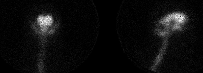

Anterior and lateral images obtained at 4 hours are shown.
View main image(ci) in a separate image viewer
View second image(ci). Anterior and right and left lateral images obtained at 24 hours are shown.
View third image(ci). Anterior and right and left lateral images obtained at 48 hours are shown.
Full history/Diagnosis is available below
CSF is primarily produced in the choroid plexus. It is transported by bulk flow into the subarachnoid space, travels throughout the spinal canal, and is ultimately primarily absorbed in the arachnoid granulations. In radionuclide cisternography radiolabeled DTPA is injected intrathecally and images are obtained at 4, 24, and 48 hours. An additional four hour image is obtained over the lumbar spine (not shown) to evaluate whether the injection was in the appropriate space. At that time tracer should be seen traveling up the spinal canal. In normal patients tracer reaches the convexities by 12 hours and the arachnoid villi in the sagittal sinus by 24 hours. Transient reflux into the lateral ventricles may be seen at 4 hours, but should clear by 24 hours. The flow in children may be somewhat faster, requiring 2, 12, and 24 hours images.
The classic (though uncommon) pattern of NPH is persistent ventricular activity without ascent over the convexities. In the mixed pattern, which is the most commonly seen, there is ventricular activity which persists up to 48 hours and subsequent ascent of tracer over the convexities. In the setting of dilated ventricles, persistent ventricular reflux allows one to diagnose a communicated hydrocephalus. If the CSF pressure is also determined to be normal, it can then be described as normal pressure hydrocephalus (NPH).
Limited data is available regarding predicting outcome following shunting. Recent onset of the symptoms of NPH (dementia, ataxia, and urinary incontinence) may be the most valuable predictor. Some (but not all) papers suggest that the typical pattern of ventricular reflux and delayed tracer ascent over the convexities predicts a favorable post-surgical outcome. Other authors have suggested that quantitative evaluation is necessary for predicting the response to shunting. The ratio of ventricular to total intracranial activity has been shown to positively correlate with response to shunting. Patients with ratios >32% improved following shunting, although some patients with lower ratios also improved following shunting.
References:
Lawrence et al., Cerebrospinal Fluid Imaging, Chapter 62 in Sandler et al., Diagnostic Nuclear Medicine (3rd ed). Williams and Wilkins Inc, 1996:1163-1173.
Larssen A, Moonen M, Bergh AC, Lindberg S, Wikkelso C. Predictive value of quantitative cisternographyh in normal pressure hydrocephalus. Acta Neurol Scand 1990;81:327-332.
Bannister R. Gilford E, Kocen R. Isotope encephalography in the diganosis of dementia due to communicating hydrocephalus. Lancet 1967;1014-1017.
Benzel EC, Pelletier Al, Levy PG. Communicating hydrocephalus in adults: prediction of outcome after ventricular shunting procedures. Neurosurgery 1990;26:665-660.
References and General Discussion of Cisternography (Anatomic field:Skull and Contents, Category:Normal, Technique, Congenital Anomaly)
Return to the Teaching File home page.