Case Author(s): Matt Jaksha, M.D. and Jerold Wallis, M.D. , 09/25/98 . Rating: #D2, #Q3
Diagnosis: Communicating Hydrocephalus
Brief history:
86 year old female with recent onset of ataxia
Images:
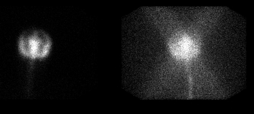
48 hour anterior and posterior images are shown
View main image(ci) in a separate image viewer
View second image(ci).
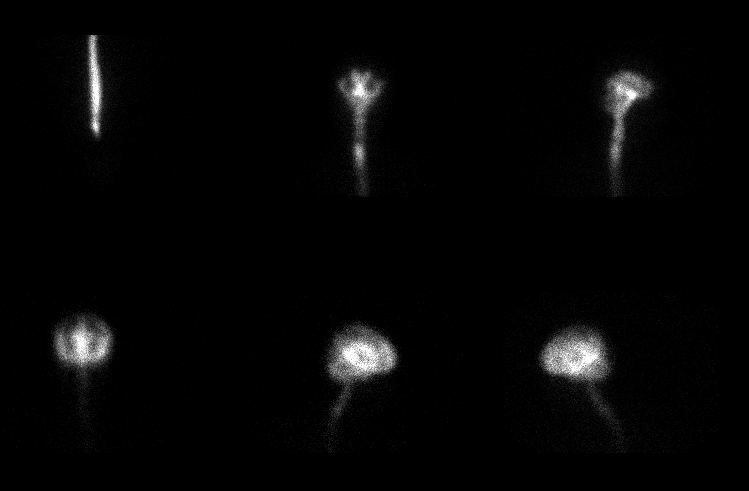
4 hour images (top row) and 24 hour images (bottom row)
Full history/Diagnosis is available below
Diagnosis: Communicating Hydrocephalus
Full history:
This 86 year old female recently developed ataxia and suffered at least one fall. 5 days prior to the cisternogram, a head CT was ordered to rule out a subdural hematoma. That demonstrated no evidence of hemorrhage. Radionuclide cisternography was requested to evaluate for normal pressure hydrocephalus.
Radiopharmaceutical:
0.46 mCi In-111 DTPA
Findings:
The 48 hour posterior image is of poor quality, with significantly increased background activity compared to the anterior image. The anterior image shows activity within the ventricular system and incomplete ascent of the tracer over the convexities. The images of the two previous days had also shown reflux into the ventricular system and delayed ascent over the convexities.
Discussion:
The marked difference in the anterior and posterior 48 hour images is the result of a technical error. The images were obtained simultaneously with a dual head camera, and the collimator for the posterior head had not been changed to a medium energy collimator. Therefore, there is significant septal penetration seen on that head. Note the star pattern typical of septal penetration.
Persistent ventricular reflux on radionuclide cisternography is abnormal. Also, usually by 24 hours and certainly by 48 hours the tracer should ascend over the convexities bilaterally as it is resorbed by the arachnoid granulations. This combination of findings is consistent with a communicating hydrocephalus (see case ci001).
Followup:
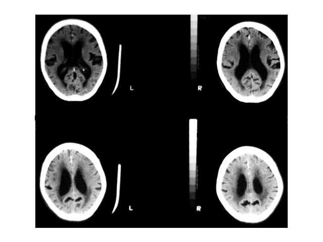
Review of the head CT from 5 days earlier demonstrates prominence of both the venticular system and the cortical sulci, particularly the sylvian fissures. There is also prominent decreased attenuation in the periventricular white matter bilaterally. In an 86 year old, these findings are nonspecific and difficult to interpret. Much of what is seen may be due simply to atrophy and white matter changes of aging. However, the pattern also corresponds well to the findings on cisternography and supports the diagnosis of a communicating hydrocephalus.
View followup image(ct).
Selected axial images from the head CT done 5 days prior
Major teaching point(s):
Higher energy isotopes need to be imaged with the proper collimator (medium or high energy) to improve resolution.
Star artifact, or streaks of increased activity radiating from a central source indicates septal penetration.
Radionuclide cisternography can be used to diagnose communicating hydrocephalus. Delayed ascent over the cerebral convexities and persistent reflux into the ventricular system is abnormal.
ACR Codes and Keywords:
References and General Discussion of Cisternography (Anatomic field:Skull and Contents, Category:Organ specific)
Search for similar cases.
Edit this case
Add comments about this case
Return to the Teaching File home page.
Case number: ci002
Copyright by Wash U MO