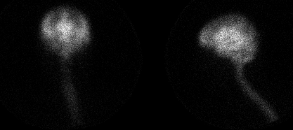
48 hr delayed cisternogram images
View main image(ci) in a separate image viewer
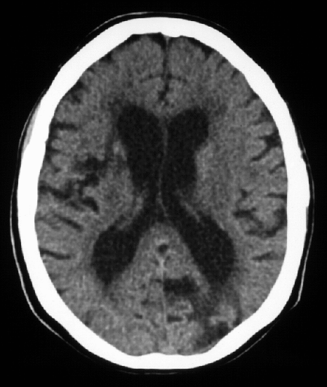
View second image(ct). Axial CT image at level of lateral ventricles
Full history/Diagnosis is available below
The total volume of CSF is about 120 mL with about one-third of this within the ventricular system and two-thirds bathing the brain and spinal cord. CSF is formed by the choroid plexus in the lateral ventricles. Normal CSF flow is through the foramen of Monroe into the third ventricle, where it exists via the aqueduct of Sylvius to the fourth ventricle. CSF then exists the ventricular system via the foramen of Luschka and Magendie to enter the basal cisterns and subarachnoid space surrounding the brain.
Normal-pressure hydrocephalus is characterized by the classic triad of dementia, ataxia, and urinary incontinence. Although the etiology of normal- pressure hydrocephalus is unknown, it remains one of the few causes of treatable pre-senile dementia. It is thought to begin with an obstruction to CSF flow either in the ventricles or basal cisterns, with resultant ventriculomegaly. The CSF pressure then slowly returns to normal with persistently enlarged ventricles. While the clinical findings are classic, the CT findings are easily confused with atrophy.The pattern of CSF flow in patients with normal-pressure hydrocephalus is abnormal and can be assessed with radionuclide cisternography.
The normal radionuclide cisternogram demonstrates activity ascending to the basal cisterns by four hours. The radiopharmaceutical will extend into the interhemispheric and Sylvian fissures forming the so-called Neptune's triumvirate. Activity should be identified over the convexities of the brain by 24 hours. Normally, there is no reflux of activity into the lateral ventricles. However, a small amount of activity which is transiently present in the lateral ventricles within the first 24 hours is within the accepted limits of normal and generally disregarded. Persistent activity within the lateral ventricles after 24 hours is virtually diagnostic in the proper clinical setting.
Treatment includes shunting of CSF flow, such as with a ventriculoperitoneal shunt. While some patients respond dramatically to treatment, the CSF flow pattern on cisternography does not predict the patientąs response to ventricular shunting. Patients with chronic normal-pressure hydrocephalus typically respond less well than those patients with acute disease. Thus, clinical data, and not imaging, determines whether a patient is to be treated.
Radionuclide cisternography does not diagnose normal-pressure hydrocephalus, per se. Rather, it diagnoses obstructive, communicating hydrocephalus in patients with a clinical suspicion of normal- pressure hydrocephalus (classic clinical triad) with ventriculomegaly on CT scan (atrophy vs NPH). The other common indication for radionuclide cisternography is detection of a CSF leak.
References:
1) Sandler PM, et al. Diagnostic Nuclear Medicine, 3rd edition, 1996. Williams and Wilkins
2) Datz, et al. Nuclear Medicine: a teaching file. 1992. Mosby Yearbook
References and General Discussion of Cisternography (Anatomic field:Skull and Contents, Category:Organ specific)
Return to the Teaching File home page.