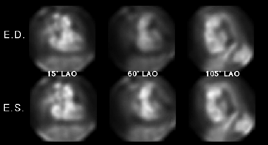

CARDIAC BLOOD POOL STUDY
End diastole (top), End systole (bottom)
View main image(ca) in a separate image viewer
Full history/Diagnosis is available below
The LV/RV stroke count ratio is decreased (0.31), suggestive of tricuspid regurgitation.
The left atrium and ventricle are normal in size and configuration. Both chambers contract normally and the calculated left ventricular ejection fraction is 55%.
The calculation of ejection fraction is (ED - ES) / (ED - background) X 100.
Right ventricular ejection fraction is particularly dependent on the afterload faced by the right ventricle. Decreased RVEF often reflects increased pulmonary artery systolic pressure or increased pulmonary vascular resistance.
The stroke count ratio (SCR) was developed to assess for the presence of valvular regurgitation. This calculation relies on the assumption that in the absence of any right sided regurgitation or intracardiac shunts, the left ventricular (LV) and right ventricular (RV) stroke volumes should be equal. In cardiac blood pool imaging, stroke counts can be substituted for stroke volumes because the two are proportional. Therefore, the change in counts between diastole and systole of the left ventricle divided by the same change in the right ventricle should equal one in the normal patient.
With left sided valvular regurgitation, the SCR is elevated (LV stroke volume increased) and with right sided regurgitation, the SCR is decreased (RV stroke count increased). This value is only valid in isolated valvular lesions because balanced bilateral lesions (ie. similar degrees of tricuspid and mitral regurgitation) may in fact cancel each other out. Also, if there are two ipsilateral valvular lesions (ie. concomittant aortic and mitral regurgitation) the ratio will reflect the sum of the lesions.
The results are dramatic! In essence, the cardiac function is now normal with complete recovery of the right ventricular function. In fact, the right atrium and ventricle are normal in size and the RVEF is now 45%.
View followup image(ca). CARDIAC BLOOD POOL STUDY (POST LUNG TRANSPLANT)
End diastole (top), End systole (bottom)
One can determine the presence of right heart enlargement quickly by merely noting the angles needed to obtain the "best septal view" and two orthogonal views (RAO and left lateral). Normally, the best septal view is obtained at approximatley 35° LAO with subsequent 45° orthogonal views at 10° RAO and 80° LAO. In this case, the initial study shows significant leftward (counterclockwise) rotation of the heart with the best septal view at 60° LAO, and orthogonal views at 15° LAO and "105° LAO" (or 75° LPO). With left heart enlargement, the heart rotates rightward and the best septal view will shift toward a more shallow LAO.
1) Chronic pulmonary embolus
2} Pulmonary fibrosis
3) Atrial septal defect with Eisenmenger's physiology (expect to also see left atrial enlargement)
4} Severe mitral stenosis (expect to also see left atrial enlargement)
5) Pulmonic stenosis (less common cause)
References and General Discussion of Cardiac Blood Pool Scintigraphy (Anatomic field:Heart and Great Vessels, Category:Misc)
Return to the Teaching File home page.