Case Author(s): Michael Roarke, M.D. and Jerold Wallis, M.D. , 2/12/96. Rating: #D2, #Q5
Diagnosis: Left ventricular pseudoaneurysm
Brief history:
66-year old woman with history
of cardiac disease who presents with worsening
congestive heart failure.
Images:

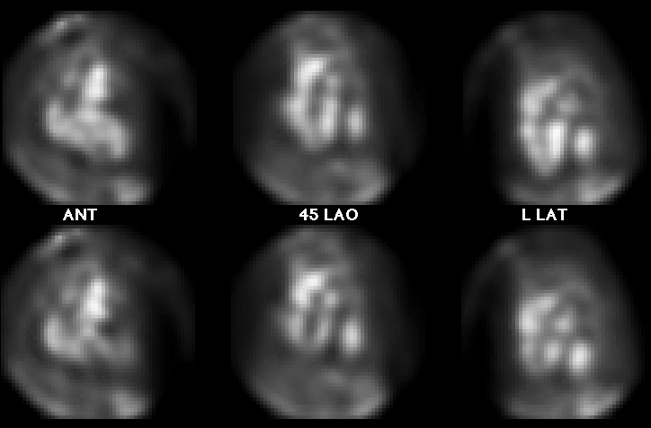
End diastole (top) and end systole (bottom)
View main image(ca) in a separate image viewer
View second image(ct).
Axial image from contrast-enhanced CT of the chest
Full history/Diagnosis is available below
Diagnosis: Left ventricular pseudoaneurysm
Full history:
66-year old woman with history
of past myocardial infarction who presents with
worsening congestive heart failure.
Radiopharmaceutical:
Tc-99m in vitro labeled
red cells
Findings:
Cardiac blood pool images obtained
at rest reveal an area of focal blood pool activity
posterior and lateral to the left ventricle. It appears
slightly more prominent on the end systolic images
than the end diastolic images. The computed
tomography images reveal a left ventricular
pseudoaneurysm corresponding to this area of
abnormal blood pool activity.
Discussion:
While true left ventricular
aneurysms represent dilatation of the myocardium
focally, usually in areas of previous infarction, left
ventricular pseudoaneurysms result from a focal
rupture of the myocardium, with the blood pool contained by the overlying
pericardium. The posterolateral location of this
pseudoaneurysm is characteristic.
Differential Diagnosis List
In addition to left ventricular
pseudoaneurysm, an alternate possibility is aneurysm of the
descending thoracic aorta. The computed tomographic
findings help to differentiate between these
possibilities.
ACR Codes and Keywords:
References and General Discussion of Cardiac Blood Pool Scintigraphy (Anatomic field:Heart and Great Vessels, Category:Misc)
Search for similar cases.
Edit this case
Add comments about this case
Return to the Teaching File home page.
Case number: ca002
Copyright by Wash U MO

