Case Author(s): Jabi Shriki, MD and Keith Fischer, MD , . Rating: #D2, #Q4
Diagnosis: Ewing's Sarcoma
Brief history:
42 year-old male with left hip pain.
Images:

Anterior and posterior delayed views from a bone scan.
View main image(bs) in a separate image viewer
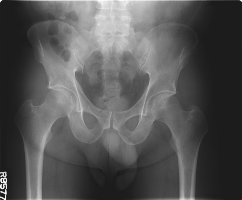
View second image(xr).
Plain AP radiograph of the pelvis.
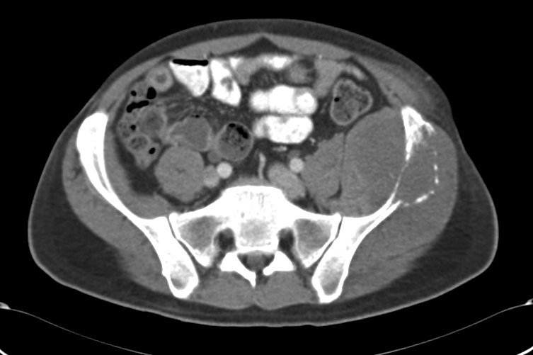
View third image(ct).
CT scan through the level of the lesion in the pelvis after IV and oral contrast administration.
Full history/Diagnosis is available below
Diagnosis: Ewing's Sarcoma
Full history:
The patient was in his usual state of health until April 2005 when he started noticing some intermittent pain in the left hip area. The pain gradually worsened with an extension toward the left iliac crest area. He sought medical attention from his primary care physician. An MRI was done at an outside hospital. This showed an interosseous process with soft tissue extension inside and outside the pelvis on his left iliac crest. Biopsy showed a malignant round cell neoplasm with morphologic and immuno histochemical features consistent with Ewing's sarcoma/peripheral neuro ectodermal tumor. Molecular studies with PCR for fusion transcript of EWS-FLI1 was also positive.
Radiopharmaceutical:
Technetium-99m Labeled MDP
Findings:
Bone scan demonstrates a subtle decrease in uptake of radiopharmaceutical in the upper portion of the left iliac crest. There is also mildly increased activity just below this.
Plain films demonstrate subtle cortical loss and bony destruction in the upper iliac crest.
Axial CT demonstrates a large soft tissue mass with lysis of the underlying bone.
Discussion:
A high index of suspicion must be maintained when bone scanning is used to evaluate for lesions which may be subtle or are predominantly lytic. Since uptake of MDP is mediated by flow and by osteoblastic activity, lytic bony lesions may be photopenic, and findings are often subtle.
Ewing's Sarcoma was first described by pathologist James Ewing in 1921. It is an uncommon, malignant bone tumor, comprising 10 to 15% of all malignant, primary bone tumors; and is also a member of the so-called "small, round, blue cell tumors" along with acute leukemia, neuroblastoma, primitive neuroectodermal tumors, Wilm's tumor, and other, more rare tumors such as small cell mesothelioma. These tumors are so named for their characteristic appearance with sheets or nests of small, tightly grouped, polygonal cells, with scant cytoplasm.
Although it's appearance may be protean, the common radiographic appearance is described as permeatively lytic, with aggressive periosteal reaction, and often a soft tissue mass. Variants are commonly described including a sclerotic pattern, and soft tissue mass effect, with little cortical bony destruction. Osteomyelitis and osteosarcoma are often differential considerations.
Treatment options are variable, but include aggressive, myeloablative chemotherapy with autologous bone marrow transplantation and radiation therapy.
Major teaching point(s):
A high index of suspicion is needed in evaluating bone scans, especially when bony lesions may be primarily lytic.
Differential Diagnosis List
Osteomyelitis and osteosarcoma are important considerations, especially in the setting of a permeative, lytic lesion. In this location, traumatic avulsion should also be considered in the appropriate clinical scenario.
ACR Codes and Keywords:
References and General Discussion of Bone Scintigraphy (Anatomic field:Skeletal System, Category:Neoplasm, Neoplastic-like condition)
Search for similar cases.
Edit this case
Add comments about this case
Return to the Teaching File home page.
Case number: bs152
Copyright by Wash U MO