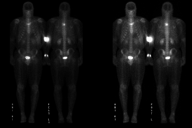

Anterior and posterior images
View main image(bs) in a separate image viewer
View second image(bs). Spot images
View third image(mr). Axial image of thoracic MRI
View fourth image(ct). CT of the spine
Full history/Diagnosis is available below
MRI shows an enhancing mass within the T4 vertebrae which obliterates the left T4-5 neural foramen and abuts the spinal canal.
CT of thoracic spine demonstrated that expansile lytic lesion within the L4 vertebral body extending into the posterior elements.
In a study of 314 people treated for LCH at the Mayo Clinic (Howarth et al, Cancer 1999;85:2278-90) 69% had single-system disease and 31% had multi-system LCH. Approximately half of those treated at Mayo were under 25 years old at diagnosis. Of people with single-system disease the system involved was bone (52%),pulmonary (lung) (40%), skin/mucous membrane (7%) and other sites (1%). Bone involvement was more common in younger patients while pulmonary involvement was mostly seen in those over 15 years old at diagnosis.
Localized pulmonary Langerhans'cell histiocytosis (also sometimes referred to as pulmonary eosinophoic granuloma) is a rare pulmonary disease that occurs predominantly in young adults.
The cause of LCH is unknown; most cases occur spontaneously.
The symptoms of LCH will depend upon which part of the body it is affecting and if the disease is affecting more than one part of the body.
Single-system LCH usually disappears on its own without any treatment. This may occur following a biopsy. In a small number of children, treatment will be needed and low-dose radiotherapy, surgery and steroids may be used. Multi-system disease is usually treated with chemotherapy and steroids.
80% of children who develop LCH will recover from it. The older the child, the higher the chance of recovery. Sometimes the disease can recur and therefore long-term followup is advised
References: Web Sites.
References and General Discussion of Bone Scintigraphy (Anatomic field:Spine and Contents, Category:Neoplasm, Neoplastic-like condition)
Return to the Teaching File home page.