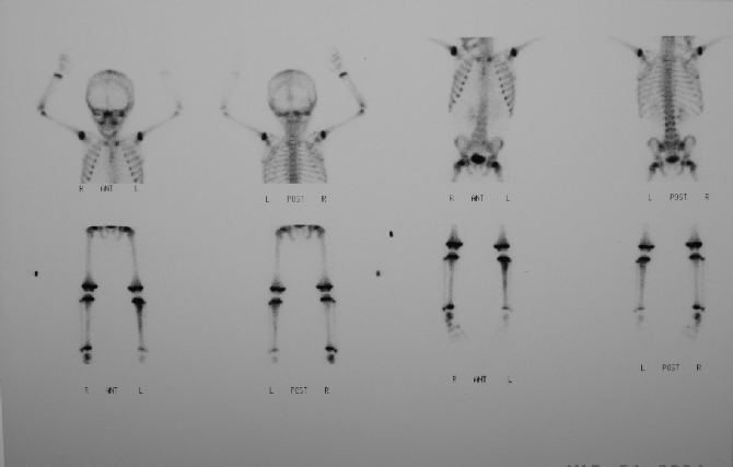
Anterior and posterior whole-body bone scintigraphy on 10/17/02
View main image(bs) in a separate image viewer
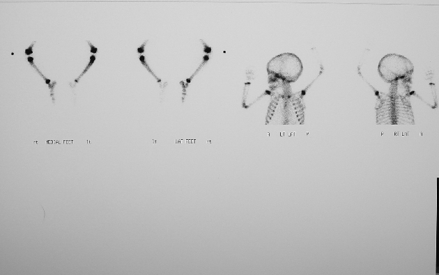
View second image(bs). Spot images
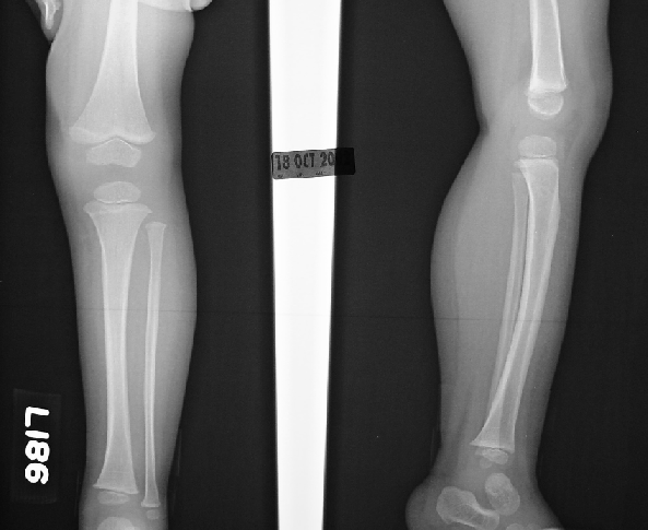
View third image(xr). Plain film of the lef leg on 10/18/04
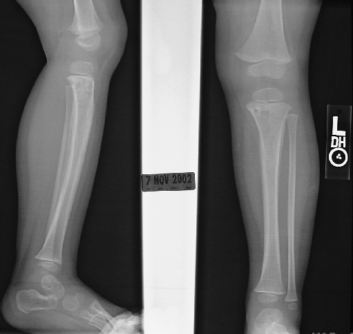
View fourth image(xr). Plain film of the left leg on 11/07/02
Full history/Diagnosis is available below
Plain films of the left tibia and fibula on 10/18/02 demonstrate a possible subtle lytic lesion in the proximal tibia and osteomyelitis cannot be excluded. Repeat films in 10-14 days may be helpful to evaluate further and to exclude fracture.
Plain films of the left tibia and fibula on 11/07/02 demonstrate an area of osteomyelitis with increasing destruction in the left proximal tibia with surrounding periosteal new bone.
In acute hematogenous osteomyelitis of children, infection involves the red marrow of the long bones as a result of the relatively slow blood flow in metaphyseal sinusoidal veins and the relative lack of phagocytes.
In acute hematogenous osteomyelitis of adults, the long bones rarely are affected because adipose tissue has replaced red marrow. Infection most often occurs in the spine with septicemia as the initiating event.
The bone scan is very sensitive for making the diagnosis. However, a positive three-phase bone scan is not specific for osteomyelitis. Fracture, tumor and Charcot joints may be three-phase positive.
Reference: Ziessman HA. Case Review of Nuclear Medicine. Mosby. 2002; 112.
References and General Discussion of Bone Scintigraphy (Anatomic field:Skeletal System, Category:Inflammation,Infection)
Return to the Teaching File home page.
Discussion:
Bone infection is usually bacterial in origin (most commonly staphylococcal) and reaches the bone by hematogenous spread, direct extension from a contiguous skin site of infection, or direct introduction by surgery or trauma.
Differential Diagnosis List
Osteomyelitis, bone tumor, fracture/osteotomy.
ACR Codes and Keywords:
Case number: bs148