Case Author(s): Zhiyun Yang, M.D. and Jerold Wallis, M.D. , 03/15/05 . Rating: #D3, #Q4
Diagnosis: Sternal metastasis from breast cancer
Brief history:
55-year-old female with breast cancer.
Images:
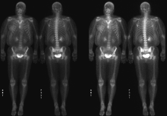
Anterior and posterior whole body images
View main image(bs) in a separate image viewer
View second image(bs).
Spot images of bone scan
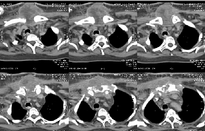
View third image(ct).
CT of the chest
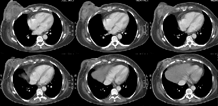
View fourth image(ct).
CT of the chest
Full history/Diagnosis is available below
Diagnosis: Sternal metastasis from breast cancer
Full history:
55-year-old female with breast cancer, status post surgery bilaterally, chemotherapy and radiation therapy. This study is to evaluate for metastatic disease.
Radiopharmaceutical:
21.0 mCi Tc-99m MDP i.v.
Findings:
Delayed whole-body scintigrams were obtained. There is mildly increased uptake seen in the right breast region which could be due to post surgical and/or radiation therapy changes or recurrent tumor. There is decreased uptake seen in the upper portion of the sternum and increased uptake in the sternal bone just beneath this area which has a corresponding finding of bony destruction on the CT scan and represents metastatic disease.
CT of the chest demonstrated that large locally invasive right breast mass seen extending through the right pectoralis major and minor to the level of the rib cage with multiple necrotic lymph nodes within the subpectoral and right axillary regions. Large suprasternal soft tissue mass likely from local spread is seen invading the sternum.
Discussion:
The sternum is a common site of local spread of breast cancer, likely from direct extension arising from internal mammary nodes. For this reason, particular attention should be paid to the sternum in the setting of breast cancer. While mild uptake at the sternomanubrial joint and the sternoclavicular (claviculomanubrial) joints is a common normal variant, abnormal uptake elsewhere in the sternum requires further investigation.
General differential diagnosis of central area of decreased uptake in bone surrounded by a rim of increased uptake:
Common: Agressive neoplasms; can also be seen in osteomyelitis and bone abscess, and previous surgery such as craniotomy.
Uncommon: Giant-cell tumor.
Rare: Coccidioidomycosis, osteoporosis circumscripta.
Followup:
Pathologic results four days after bone scintigraphy:
A. BREAST, LEFT, MODIFIED RADICAL MASTECTOMY
- Invasive ductal adenocarcinoma, poorly differentiated.
B. SOFT TISSUE, RIGHT AXILLARY MASS, EXCISION
- Metastatic adenocarcimoma, poorly differentiated
C. CHEST WALL, ANTERIOR, BIOPSY
- Metastatic adenocarcimoma, poorly differentiated
D. SOFT TISSUE, ANTERIOR CHEST WALL MASS
- Metastatic adenocarcimoma, poorly differentiated, consistent with breast primary
ACR Codes and Keywords:
References and General Discussion of Bone Scintigraphy (Anatomic field:Skeletal System, Category:Neoplasm, Neoplastic-like condition)
Search for similar cases.
Edit this case
Add comments about this case
Return to the Teaching File home page.
Case number: bs147
Copyright by Wash U MO