Case Author(s): Delphine Chen, MD and Henry Royal, MD , 08/26/04 . Rating: #D3, #Q5
Diagnosis: Rhabdomyolysis
Brief history:
18 yo. male with darkening urine, worsening muscle pain, and decreasing urine output over the past 3 days after one day of intense physical exercise.
Images:

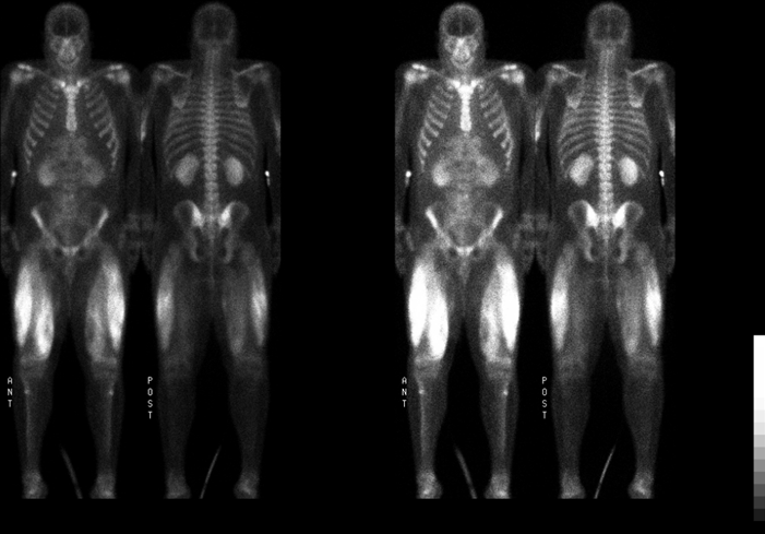
Whole body delayed images shown.
View main image(bs) in a separate image viewer
View second image().
Spot images.
Full history/Diagnosis is available below
Diagnosis: Rhabdomyolysis
Full history:
18 yo. male who presented from an OSH with rhabdomyolysis and acute renal failure. He reported a 3-day history of darkening urine, increasing muscle pain, and decreasing urine output after one day of intense physical training. Patient reported taking 2 tablets of KCl after finishing training and multiple tablets of ibuprofen to control muscle pain with no relief.
Whole body bone scintigraphy was ordered to identify the extent of involvement of rhabdomyolysis as there was concern about the development of compartment syndrome in the affected muscles.
Radiopharmaceutical:
20.8 mCi Tc-99m methylene diphosphonate i.v.
Findings:
There is intense uptake in the thighs bilaterally, particularly in the vastus lateralis and medialis muscles. In the vastus medialis muscles bilaterally, there is a central area of decreased activity which suggests more extensive necrosis in these areas. There is also moderate uptake in the triceps bilaterally and mild uptake in the rectus muscle superiorly.
Additionally, uptake in the kidneys is higher than normal, and no bladder activity is seen, consistent with the patient's acute renal failure.
Discussion:
Tc-99m MDP can accumulate in soft tissue as a result of a number of different conditions and is probably the result of soft tissue calcification or infarction.
In rhabdomyolysis, the mechanism of uptake is thought to be related to muscle cell injury and subsequent infarction, which disrupts the local calcium-phosphate balance, causing Tc-99m to be deposited in the areas of involvement. However, inflammation of the affected muscles could also increase flow and uptake of the tracer. Bone scintigraphy can be used to identify all involved muscle groups and the degree of involvement, as it was in this case.
Causes of rhabdomyolysis, besides intense physical exercise, include crush injury due to trauma and adverse reactions to drugs, such as statins, or drugs of abuse, such as cocaine.
References:
Mettler FA and Guiberteau MJ. Essentials of Nuclear Medicine, 4th ed. Philadelphia, W.B. Saunders, 1998, p299.
Followup:
On admission, the patient's creatinine was 4.2 mg/dl. Creatine kinase was 261000 IU/L (normal range 30-200 IU/L) and myoglobin was 21900 ng/ml (normal range 0-110 IU/L), which both peaked the following day. Hospital course was complicated by hypertension which was difficult to control. Patient did not develop compartment syndrome and eventually recovered without need for dialysis, with myoglobin and serum creatine kinase levels trending towards normal, and was discharged home on medications for persistent hypertension.
Differential Diagnosis List
Other possibilities for muscle uptake of Tc-99m MDP include:
-Myositis ossificans
-Polymyositis
-Electrical burns
ACR Codes and Keywords:
References and General Discussion of Bone Scintigraphy (Anatomic field:Skeletal System, Category:Metabolic, endocrine, toxic)
Search for similar cases.
Edit this case
Add comments about this case
Return to the Teaching File home page.
Case number: bs144
Copyright by Wash U MO

