Case Author(s): Feiyu Xue, M.D., Ph.D. and Jerold Wallis, M.D. , 6/28/04 . Rating: #D2, #Q3
Diagnosis: Non-ossifying fibroma
Brief history:
15-year-old male with a history of right leg swelling and pain
Images:

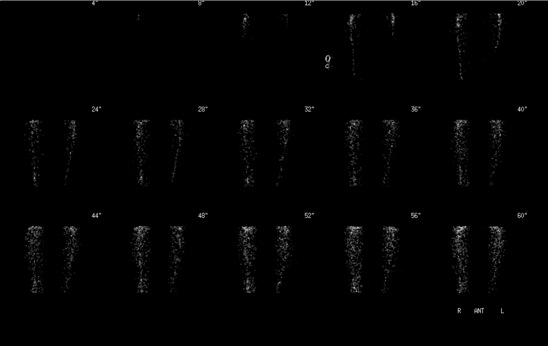
Bone scintigraphy, angiographic phase
View main image(bs) in a separate image viewer
View second image(bs).
Bone scintigraphy, immediate static
View third image(bs).
Bone scintigraphy, delayed
Full history/Diagnosis is available below
Diagnosis: Non-ossifying fibroma
Full history:
15 year-old male with a history of right leg swelling and pain. Although he sprained his ankle some time ago, he has had no recent injury. This study is to evaluate for possible bone lesion or fracture.
Radiopharmaceutical:
18.6 mCi MDP i.v.
Findings:
A limited examination of the right leg and foot was performed consisting of radionuclide angiography, immediate post-injection images, and delayed magnification images. There is mildly increased blood flow seen in the distal right leg on radionuclide angiographic images. Focally increased activity is noted in the right distal tibia on both immediate post-injection images and delayed images, which has a corresponding bone lesion in the right distal tibia on plain radiographs (see follow-up image below).
Discussion:
Non ossifying fibroma (NOF) is a benign bone lesion occurring in the developing skeleton. Peak occurrence is in those aged 10-15 years, with decreasing incidence in adolescents. The overall age range of patients is reported to be 3-20 years. NOF almost never appear for the first time in adults and they are thought to persist from childhood if they are present in adults. Typically, NOFs are asymptomatic and detected only incidentally on radiographs obtained for other reasons. Approximately 90% of cases of both lesions involve the tubular long bones. Common sites include the distal femur, the proximal and distal tibia; most lesions occur around the knee. On bone scintigraphy, Minimal-to-mild accumulation MDP occurs and is indicative of a benign process. The intensity of the uptake is typically less than that of active bone lesions (fracture, infection, and neoplasm). In a child or adolescent, an eccentric region of uptake located a short distance from the physis of a tubular bone with the appropriate corresponding plain radiographic findings is diagnostic for NOF. In children, this finding can be masked by the proximity of the lesion to the distal femoral epiphyseal plate, which itself has marked tracer uptake.
Reference:
Marks KE, Bauer TW. Fibrous tumors of bone. Orthop Clin North Am. 1989 Jul;20(3):377-93.
View followup image(xr).
Radiography of the distal right tibia and fibula
Major teaching point(s):
Increased uptake on bone scan is non-specific. Benign lesions like non-ossifying fibroma usually have uptake less than that of active lesions (fracture, infection, and neoplasm. When abnormalities are seen on bone scan, correlation with plain radiographs is useful.
Differential Diagnosis List
Bone tumor,
Osteomyelitis,
Fracture
ACR Codes and Keywords:
References and General Discussion of Bone Scintigraphy (Anatomic field:Skull and Contents, Category:Neoplasm, Neoplastic-like condition)
Search for similar cases.
Edit this case
Add comments about this case
Return to the Teaching File home page.
Case number: bs140
Copyright by Wash U MO

