Case Author(s): Yonglin Pu, MD. PhD, Keith Fischer,MD. , 03/23/03 . Rating: #D2, #Q4
Diagnosis: Multiple Stress Fractures
Brief history:
A 17-year-old male has a three-week history of right lower extremity pain. The patient is a long distance runner.
Images:

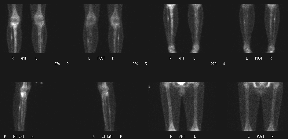
Bone scintigraphy of lower extremities, delayed images
View main image(bs) in a separate image viewer
View second image(bs).
Bone scintigraphy of lower extremities, radionuclide angiography and immediate static images
View third image(xr).
Plain X-ray of right lower leg
Full history/Diagnosis is available below
Diagnosis: Multiple Stress Fractures
Full history:
A 17-year-old male has a three-week history of right lower extremity pain. The patient is a long distance runner averaging more than 3 miles per day. Please evaluate for possible stress fracture.
Radiopharmaceutical:
16.0 mCi Tc-99m MDP i.v.
Findings:
A limited examination of the lower extremities was performed consisting of a radionuclide angiography, immediate post-injection images, and delayed images. The study was compared to the plain radiographs of the right tibia and fibula performed 4 days previously.
There is increased flow and increased uptake on the immediate post-injection images in the right upper tibia. On the delayed images, there is focally increased activity in the right proximal tibial plateau, the proximal third of the right fibula, and multiple foci of increased activity in the right tibial diaphysis. These findings are consistent with stress fractures in different phases of healing. There is focally increased activity in the mid left fibula and proximal third of the left tibia, also consistent with stress fractures. Some of the increased uptake correlates with solid periosteal reaction seen along the distal fibular diaphysis and the proximal tibial diaphysis on comparison X-ray images.
Discussion:
Stress fractures occur in normal bone that undergo repeated abnormal stress. In this case the repeated and abnormal stess is secondary to running. Bone scintigraphy normally becomes positive at the same time or sooner than plain films and has a low false-negative rate. The most common sites of stress fracture are the femoral neck and tibia. Less commonly, the femoral diaphysis may be involved.
References:
Donohoe K. Selected topics in orthopedic nuclear medicine. Orthop clin N Am. 29, 85-101. 1998.
Holder L. Bone scintigraphy in skeletal trauma. Rad clin N Am. 31, 739-781. 1993.
Wilcox J, et al. Bone scanning in the evaluation of exercise-related stress injuries. Rad. 123, 699-703. 1977.
Differential Diagnosis List
This is a very specific pattern for stress fractures. "Shin splints" which occur in long distance runners typically occur over a longer lenth of the tibia at the insertion of the posterior tibialis muscle.
ACR Codes and Keywords:
References and General Discussion of Bone Scintigraphy (Anatomic field:Skeletal System, Category:Effect of Trauma)
Search for similar cases.
Edit this case
Add comments about this case
Return to the Teaching File home page.
Case number: bs133
Copyright by Wash U MO

