Case Author(s): Jayson R. St. Jacques, M.D. and Barry A. Siegel, M.D. , . Rating: #D3, #Q3
Diagnosis: Left shoulder osteomyelitis
Brief history:
49-year-old woman with a history of breast cancer
Images:

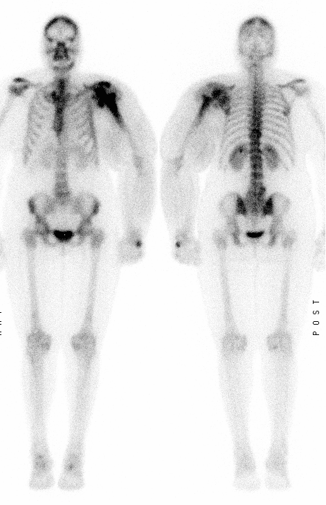
Anterior and posterior whole-body bone images are shown.
View main image(bs) in a separate image viewer
View second image(xr).
Anteroposterior radiograph of left arm.
View third image(mr).
Coronal T1-weighted fat-saturated MR image of left shoulder.
View fourth image(mr).
Axial T1-weighted post-gadolinium MR image of left shoulder.
Full history/Diagnosis is available below
Diagnosis: Left shoulder osteomyelitis
Full history:
49-year-old woman with a history of left breast cancer treated 10 years ago with mastectomy, followed by radiation and chemotherapy. She now presents with dizziness, falling, and left shoulder pain. She also has swelling and weakness in the left arm.
Radiopharmaceutical:
20.7 mCi Technectium-99m MDP i.v.
Findings:
Bone scintigraphy demonstrates increased activity in the left scapula, clavicle, and proximal humerus, centered around the shoulder joint. There is decreased activity in the left humeral head, consistent with bone destruction. The left upper extremity is markedly enlarged, likely due to lymphedema or venous compression. Notably, although there are foci of mildly increased activity in the lower thoracic spine and the lower lumbar spine, likely due to degenerative disease, there are no other lesions suggesting metastatic disease.
The radiograph shows a destructive lesion of the left proximal humerus and a large soft tissue mass. The MR images also show a destructive lesion centered in the left proximal humerus (completely destroying the humeral head) with an associated soft tissue component.
Followup:
Ultrasound-guided biopsy documented infection secondary to Staphylococcus aureus. Open exploration confirmed the osteomyelitis of the humerus with extension into the glenhumeral joint.
Differential Diagnosis List
The differential diagnosis for this extensive destructive lesion includes metastatic breast carcinoma, a radiation-induced sarcoma, and infection. The apparent involvement on both sides of the joint favors infection or a soft-tissue tumor rather than an primary bone tumor or an osseous metastasis.
ACR Codes and Keywords:
References and General Discussion of Bone Scintigraphy (Anatomic field:Skeletal System, Category:Inflammation,Infection)
Search for similar cases.
Edit this case
Add comments about this case
Return to the Teaching File home page.
Case number: bs131
Copyright by Wash U MO

