Case Author(s): Paul Colomb, M.D. and Barry A. Siegel, M.D. , 12/10/01 . Rating: #D2, #Q4
Diagnosis: Incorrect pulse-height analyzer window setting
Brief history:
73-year-old woman with low back pain
Images:
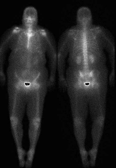
Anterior and posterior whole-body bone images
View main image(bs) in a separate image viewer
View second image(bs).
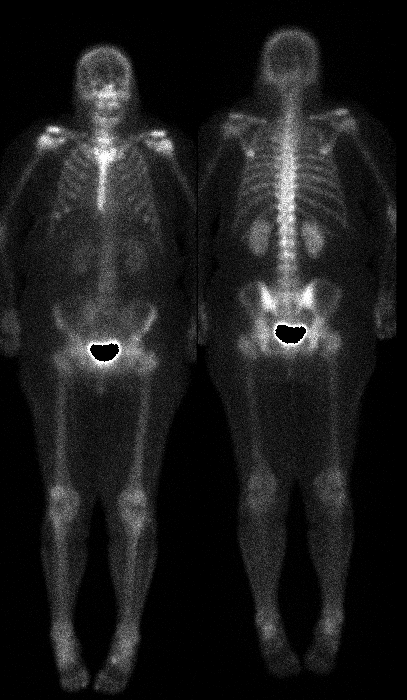
Repeat anterior and posterior whole-body bone images obtained immediately after the first set.
Full history/Diagnosis is available below
Diagnosis: Incorrect pulse-height analyzer window setting
Full history:
73-year-old woman with low back pain and known degenerative disk disease. She has no prior history of malignancy.
Radiopharmaceutical:
20.5 mCi Tc-99m MDP i.v.
Findings:
The initial whole-body bone images are of poor technical quality. There is poor resolution of skeletal detail and poor contrast. The second set of images, obtained after correction of the problem, are of good technical quality and demonstrate no abnormalities.
Discussion:
The initial whole-body bone images were obtained with the camera's pulse-height-analyzer window incorrectly set for the photopeak of Co-57. This is a fairly common technical artifact. Each day, the camera uniformity is checked with a flood source containing Co-57, a radionuclide with a long physical half-life (271.8 days) that decays by electron capture and emits a primary gamma photon at 122 keV. The Co-57 window setting wasn't changed to that for Tc-99m (140 keV) before whole-body bone imaging was performed. The image obtained with the window centered at 122 keV is of poor quality because a large fraction of scattered radiation was included in the image. This results in both degraded resolution and reduced contrast.
Poor resolution on a whole-body bone image might also result from excessive image noise (e.g., because too little Tc-99m MDP was administered or because the image was obtained at too high a scan (table) speed. In such a case, more image mottle would have been apparent than in this case. Also, poor resolution could be due to an excessive distance from the patient to the collimator, but contrast would be less degraded in that circumstance.
Major teaching point(s):
During image interpretation, it is essential to assess for technical artifacts that could be degrading image quality.
Differential Diagnosis List
Excessive image noise (low administered activity, too rapid scan speed).
Excessive patient-to-collimator distance.
ACR Codes and Keywords:
References and General Discussion of Bone Scintigraphy (Anatomic field:Skeletal System, Category:Other(Artifact))
Search for similar cases.
Edit this case
Add comments about this case
Return to the Teaching File home page.
Case number: bs129
Copyright by Wash U MO