Case Author(s): Jeffrey W. Gerstel, D.O. and Keith Fischer,M.D. M.D. , 1/12/02 . Rating: #D3, #Q3
Diagnosis: Myo-fasciitis left anterior thigh
Brief history:
6-year-old male with a bicuspid aortic valve and bacterial endocarditis. The patient has symptoms of left hip and proximal left thigh pain.
Images:
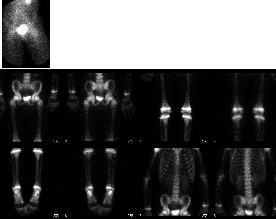
Immediate static and delayed whole body bone scan
View main image(bs) in a separate image viewer
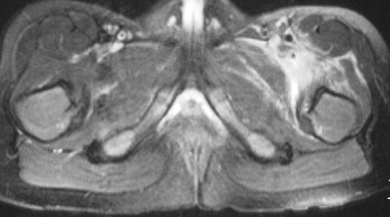
View second image(mr).
An axial slice through the pelvis - fat suppressed turbo spin echo T2
Full history/Diagnosis is available below
Diagnosis: Myo-fasciitis left anterior thigh
Full history:
6-year-old male with known bicuspid aortic valve. He presented to the hospital with fevers and left hip pain. Blood cultures grew streptococcus. He was eventually diagnosed with endocarditis and placed on intravenous antibiotics. An echocardiogram demonstrated severe aortic insufficiency. The patient refused to walk beasuse of left hip pain, but plain radiographs of the pelvis and an ultrasound of the hips were normal. The patient eventually developed a skin rash of the proximal anterior left thigh. A bone scan and an MRI were requested for further evaluation of the patient's left hip pain.
Radiopharmaceutical:
4.9mCi Tc-99m MDP i.v.
Findings:
On the immediate static image of the pelvis and proimal thighs, there is increased activity noted in the soft tissues of the left hip/left thigh. The delayed whole-body bone scan demonstrate normal distribution of activity throughout the skeleton. These findings represent a focal area of soft tissue hyperemia without evidence for osteomyelitis. An axial MRI slice through the pelvis, which was performed a day later than the bone scan, shows abnormal T2 hyperintensity surrounding the neurovascular bundle in the left hip/proximal left thigh. The greatest extent is between the satorius and the adductor muscles. The bone marrow signal is normal. There is no focal fluild collection or hip joint effusion. These findings are consistent with a myo-fasciitis of the left anterior thigh without evidence for an abscess.
Discussion:
Nuclear medicine has an important role in aiding the diagnosis of musculoskeletal infections. Unlike plain radiographs which tend to be not very sensitive for the detection of infection, a three-phase bone scan is both sensitive and specific for osteomyelitis in an otherwise normal bone in a pediatric patient. A positive bone scan with all three phases (angiographic, immediate static and delayed bone scan) can have a sensitivity that exceeds 95%. In the case above, only the first two phases of a three phase were positive, thus virtually excluding the diagnosis of osteomyelitis. When the first two phases of a three-phase bone scan are positive, soft tissue infection is the most likely diagnosis.
Other diagnostic tools have also been helpful in the evaluation of musculoskeletal infections. Magnetic Resonance Imaging (MRI) has demonstrated a high sensitivity and good anatomic correlation. Ultrasound has been helpful in detecting fluid collections in joint and soft tissues and may be used as a guide for aspirations and drainages. Labeled leukocyte imaging in combination with a bone or bone marrow scan have aided in the identification of infection especially in cases where orthopedic hardware or a diabetic foot may complicate the evaluation for infection.
Followup:
Since there was no evidence for an osteomyelitis and the patient was already on intravenous antibiotics for endocarditis, no further treatment was initiated for his myo-fasciitis. The patient eventually had a Ross procedure and his post-operative course was uneventful. The patient was discharged to home with intrvenous antibiotics.
Major teaching point(s):
1) Gold RH, Hawkins RA and Katz RD. Bacterial osteomyelitis: findings on plain radiography, CT, MR, and scintigraphy. AJR Am J Roentgenol 1991 Aug; 157(2):356-70.
2) Kothari N., Pelchovitz DJ, and Meyer JS. Imaging of musculoskeletal infections. Radiol Clin North Am 2001 Jul;39(4):653-71.
3) Mazur JM, Ross G, Cummings J, Hahn GA Jr, McCluskey WP. Usefulness of magnetic resonance imaging for the dignosis of acute musculoskeletal infections in children. J pediatr Ortho 1995 Mar-Apr; 15(2):144-7.
4) Mettler, Fred A. and Guiberteau, Milton J.. Essentials of Nuclear Meicine Imaging 4th ed.. WB. Saunders Company. 1998.
5)Palestro CJ and Torees MA. Radionuclide imaging in orthopedic infections. Semin Nucl Med 1997 Oct;27(4)334-45.
6) Sammak B, Abd El Bagi M, Al Shahed M, Hamilton D, Al Nabulsi J, Youssef B, Al Thagafi M. Osteomyelitis: a review of currently used imaging techniques. Eur Radiol 1999;9(5):894-900.
Differential Diagnosis List
1) cellulitis
2) electrical burns
3) surgical wound
4) urine contamination
5) contusion
ACR Codes and Keywords:
- General ACR code: 42
- Skeletal System:
44. "PELVIS, HIP JOINT, THIGH exclude: sacrum (33.)"
References and General Discussion of Bone Scintigraphy (Anatomic field:Skeletal System, Category:Inflammation,Infection)
Search for similar cases.
Edit this case
Add comments about this case
Return to the Teaching File home page.
Case number: bs128
Copyright by Wash U MO