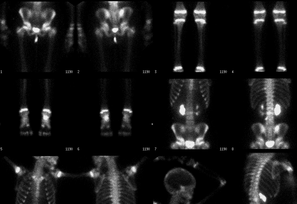
Whole body bone scintigraphy (08/22/01)
View main image(bs) in a separate image viewer
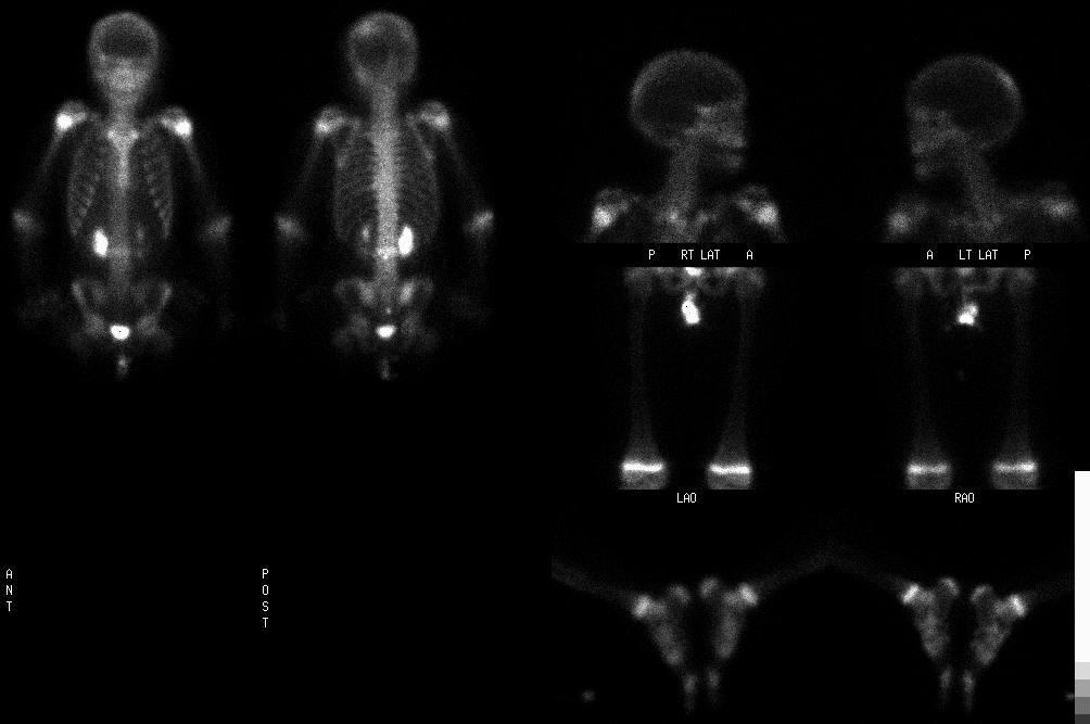
View second image(bs). Whole body bone scintigraphy (09/28/01)
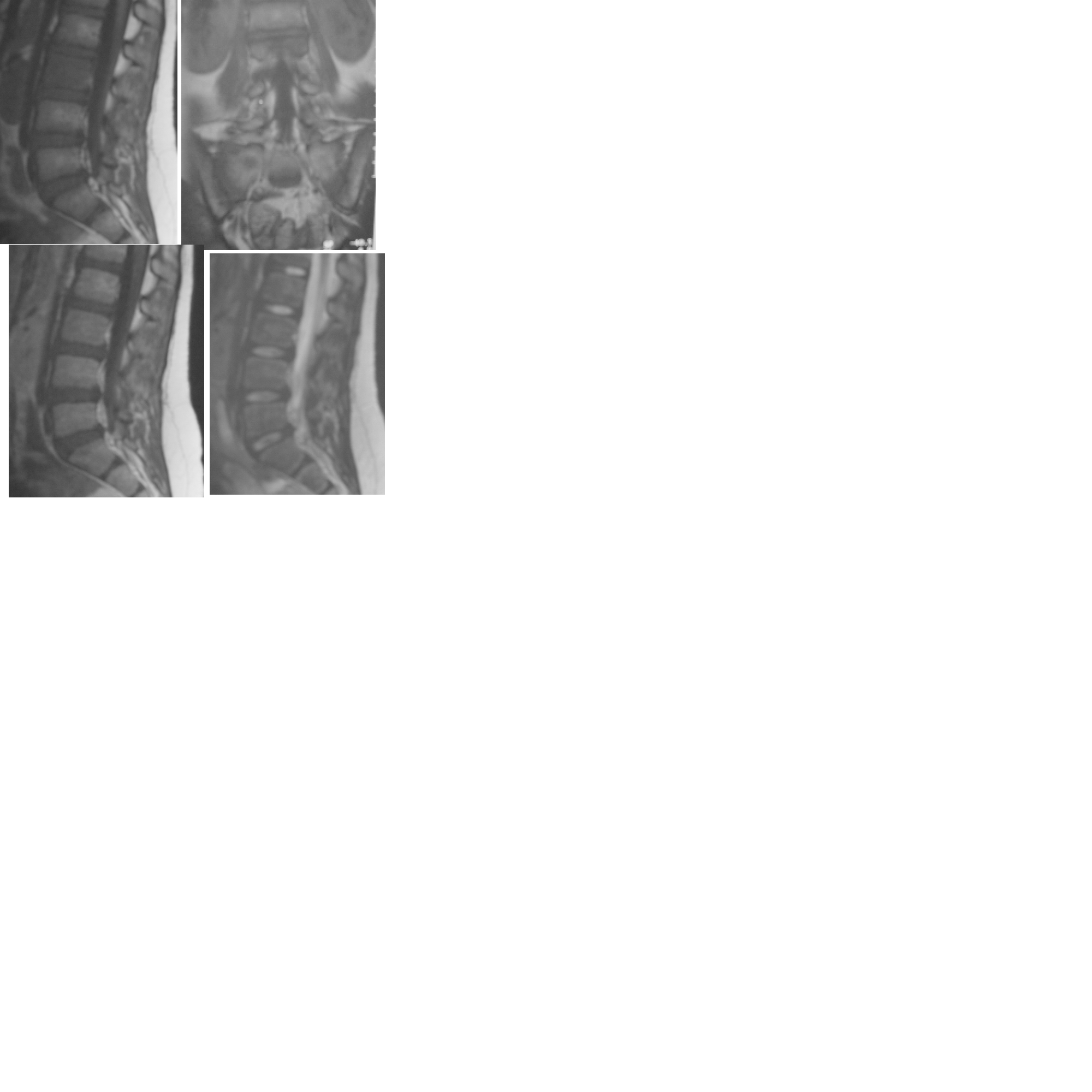
View third image(mr). Upper row: Sagittal T1WI and coronal T1WI SE MR images;Bottom row:sagittal Gd enhancement T1WI and sagittal T2WI SE MR images (08/24/01)
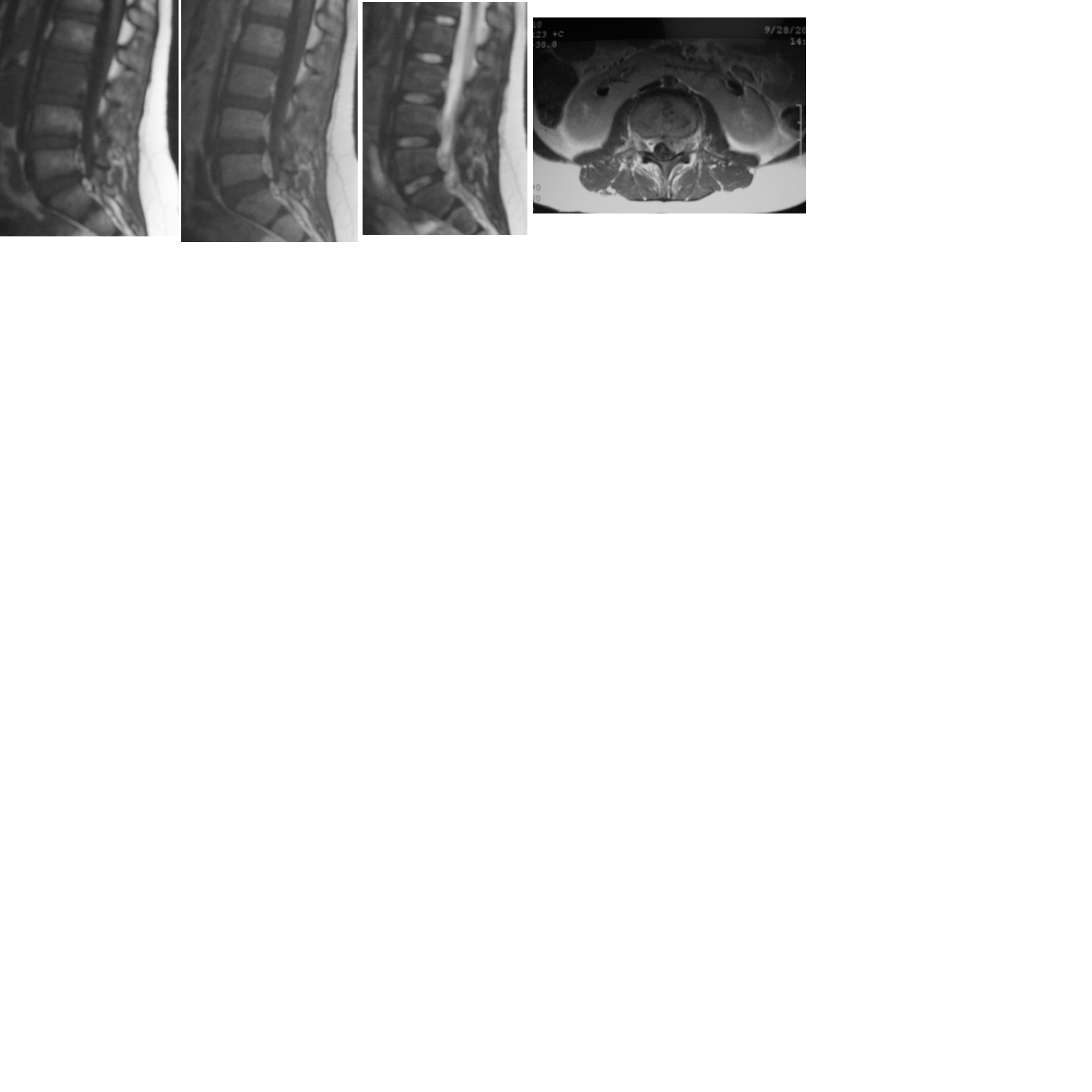
View fourth image(mr). Upper row: Sagittal T1WI and T1WI Gd enhanced, and T2WI SE images;Bottom row:Axial Gd enhanced T1WI SE MR image through L3(09/28/01).
Full history/Diagnosis is available below
BONE SCINTIGRAPHY (09/28/01): There are focally increased activity in the following regions: left parietal-occipital junction, the head of the left 9th rib at the costovertebral junction, posterior segment of the right 6th rib, L3 and L4 vertebrae, right sacrum and left calcaneus. Compared with the prior bone scintigraphy dated 08/22/01 (1st image), there is some improvement in the right posterior 6th rib and left calcaneal lesions, but there is subtle worsening in the L3 lesion, and no significant change in the left posterior 9th rib, L4 and right sacrum lesions.
MRI OF THE LUMBAR SPINE WITHOUT AND WITH GADOLINIUM ENHANCEMENT(08/24/2001). There are low T1 signal lesions involving the majority of the L3 vertebral body and the left portion of the L4 vertebral body. These lesions show heterogeneous enhancement after contrast administration. There is no associated epidural soft tissue mass. There is also a hypointense lesion in the right side of sacrum on T1-weighted images, measuring 1.5 cm in diamter withmild enhancement.
MRI OF THE LUMBAR SPINE WITHOUT AND WITH GADINIUM ENHANCEMENT(9/28/2001). An abnormal marrow signal is seen in L3 and L4, which is hypointense signal on T1 weighted images and enhances following I.V. contrast. There is epidural extension and bilateral pedicle involvement in the area posterior to the L3 vertebral body, which results in indentation of the left thecal sac (not shown). However, there is no loss of vertebral body height is identified. The intervertebral disks are normal in appearance.
LUMBOSCRAL SPINE (09/13/01) was normal radiographically.
LEUKOCYTE SCINTIGRAPHY(10/03/01):In the region of L3, there is decreased uptake of In-111 labeled white blood cells corresponding to the increased uptake of tracer demonstrated on the bone scintigraphy and L3 with abnormal MRI signal on the MRI images. There were no other areas of abnormally increased uptake of In-111 labeled white blood cells to correspond to the bone scintigraphic abnormalities.
CT SCAN WITHOUT AND WITH CONTRAST ENHANCMENT (08/23/01): There is a round 7-mm low-attenuation focus in the right hepatic lobe, which moderately enhances.
Cat-scratch disease caused by B. henselae is relatively common cause of chronic regional lymphadenopathy in children and young adults. A history of the exposure to cats can usually be elicited. One or more non-pruritic papules usually develop at the site of the cat scratch, followed by regional lymphadenopathy in about 3 to 4 weeks. Patient can present with fever or without fever. Disseminated involvement is rare and usually occurs in immuno-compromised patients and presents with bacillary angiomatosis, abcess formation, osteomylitis, or encephalitis (2).
The diagnosis of cat-scratch disease is based on clinical and enzyme immunoassay serology or polymerase chain reaction of aspirates. The use of imaging studies have been reported in the diagnosis of either localized lymphadenopathy or multifocal osteomyelitis(2-8).
For the lymphadenopathy, plain X-ray films may show soft tissue swelling or focal soft tissue mass. Ultrasound can demonstrate enlarged lymph nodes, which are often hypoechoic due to necrosis. On contrast enhanced CT, the lymph nodes are enhanced in their peripheries and not enhanced in their center because of central necrosis. On the MRI images, the lymph nodes are intermediate in signal intensity with irregular margins on T1-weighted images. Extensive edema can also be identified on the T1- and T2-weighted images. The necrotic center in the lymph nodes can also be identified as non-enhanced center on gadolinium contrast enhanced images. On Bone scintigraphy, the lymph nodes can have increased flow on arterial phase, increase blood volume on immediate static images. Delayed images can show increased activity in the adjacent bone, if it is affected by the inflammation in the lymph nodes(3).
As for the diagnosis of multifocal osteomyelitis with imaging modalities, there are a few case reports using MRI, CT and bone scintigraphy. In two case reports, T1-weighted MRI images showed para-vertebral soft tissue mass or multifocal regions of abnormally decreased signal intensity on T1-weighted SE images and abnormally hyperintensity in spine on T2-weighted images. Contrast enhancement T1-weighted images showed increased signal intensity (4-5). In another patient, CT images of chest and abdomen showed enhanced paraspinous fluid colletion adjacent to and arise from ribs. Delayed Bone scintigraphy demonstrated multifocally increased uptake in the skeleton, like in the ribs, right iliac crest and acetabulum (6-8).
References: 1. Lucy Stuart Tompkins. Bartonella Infections, Including Cat-Scratch Disease. In www.harrisonsonline.com 2. Struk DW, Munk PL, Lee MJ, Ho SG and Worsley DF. Imaging of soft tissue infection. Radiologic Clinics of North America.2001;39 (2):277. 3. Walter JN, Nimityongskul P. Positive bone scan in cat-scratch disease. The Pediatric Infectious Disease Journal 1987;6(10):944-945. 4. Robson JM, Harte GJ, Osborne DR, McCormack JG. Cat-scratch disease with paravertebral mass and osteomyelitis. Clin Infect Dis. 1999 Feb;28(2):274-278. 5. Ratner LM, Kesack A, McCauley TR, Disler DG Disseminated Bartonella henselae (cat-scratch disease): appearance of multifocal osteomyelitis with MR imaging. AJR Am J Roentgenol 1998 Oct;171(4):1164-5 6. Modi SP, Eppes SC, Klein JD. Cat-scratch disease presenting as multifocal osteomyelitis with thoracic abscess. Pediatr Infect Dis J 2001 Oct;20(10):1006-1007. 7. Breitschwerdt E, Kordick D. Bartonellosis. J Am Vet Med Assoc 1995;206:1928-1931. 8. Adal K, Corkerel C, Petri W. Cat Scratch disease, bacillary angiomatosis and other infections due to rocholimea. N. Engl J Med 1994;330;1509-1515.
References and General Discussion of Bone Scintigraphy (Anatomic field:Skeletal System, Category:Inflammation,Infection)
Return to the Teaching File home page.