Case Author(s): Yungao Ding, MD, PhD and Farrokh Dehdashti, MD , 05/07/2001 . Rating: #D., #Q.
Diagnosis: Osteoblastoma
Brief history:
34 year old woman with persistent neck pain
Images:
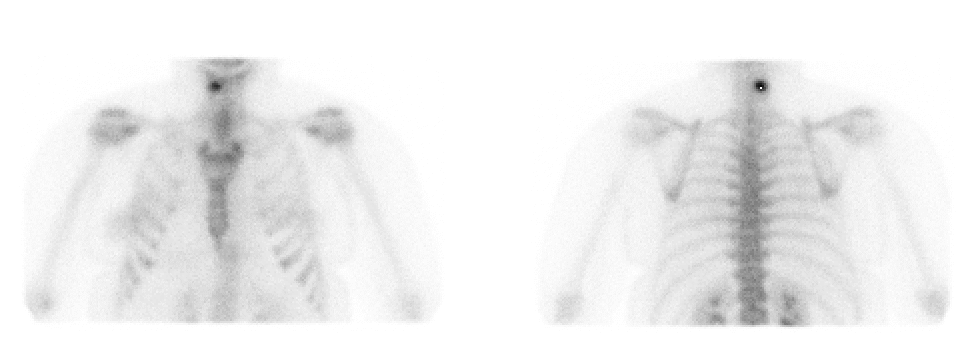
Anterior and posterior delayed planar images of cervical spine
View main image(bs) in a separate image viewer
View second image(bs).
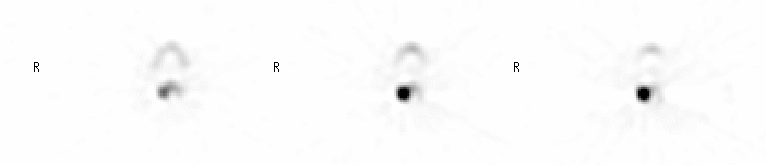
Transaxial SPECT images of cervical spine at C4-5
View third image(bs).
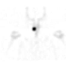
Cronal SPECT image of the cervical spine
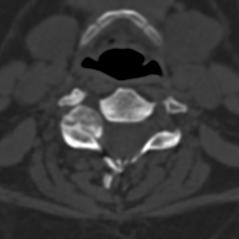
View fourth image(ct).
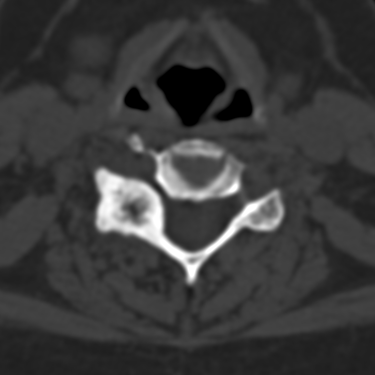
CT of the cervical spine at C5
Full history/Diagnosis is available below
Diagnosis: Osteoblastoma
Full history:
34 year old female with a history of persistent neck and clinical suspicion of osteoblastoma
Radiopharmaceutical:
20.8 mCi Tc-99m MDP, i.v.
Findings:
A focal intense uptake of the radiotracer at region of the right C4-5 facet joint as demonstrated on both planar and SPECT images classically seen in osteoblastoma/osteoid osteoma.
CT of the cervical spine showed expansile and sclerotic changes centered on the junction of the right superior facet, transverse process and lamina of the C-5. The radiolucent center of the lesion likely represented the nidus of the osteoblastoma, which was later confirmed on the surgical specimen. There was no associated soft tissue mass.
Discussion:
Osteoblastoma is rare benign tumor with unlimited growth potential and capability of malignant transformation. Lesions less than 1.5 cm are called osteoid osteoma. Osteoblastoma classically shows intense uptake of radiotracer on bone scan. There is 10% recurrence after surgical excision.
Followup:
CT and MRI of the cervical spine were performed prior to bone scan. However, the reports of CT and MIR stated that the changes were more consistent with severe degenerative arthropathy at the right C4-C5 facet joint. There was erosion of the facet joint surface, expansile and sclerotic changes on both sides of the joint although the chnages were more severe at the side of C-5. There was a small disk protrusion into the right C4-C5 neural foramen, marked disk space narrowing and straightening of the normal cervical lordosis at C4-C5. But the findings are so asymmetrical, a underlying bony lesion was strongly suspected.
The patient subsequently underwent surgical resection of the right posterior element of C-5, which showed a osteobalstoma.
View followup image(ct).
Transaxial CT image through C4-C5 facet joint
Major teaching point(s):
When severe degenerative changes of the spine are dramatically asymmetrical, an underlying bone lesion should be suspected, especially in young patients unless there is history of trauma, surgery, etc.
ACR Codes and Keywords:
References and General Discussion of Bone Scintigraphy (Anatomic field:Spine and Contents, Category:Neoplasm, Neoplastic-like condition)
Search for similar cases.
Edit this case
Add comments about this case
Return to the Teaching File home page.
Case number: bs126
Copyright by Wash U MO