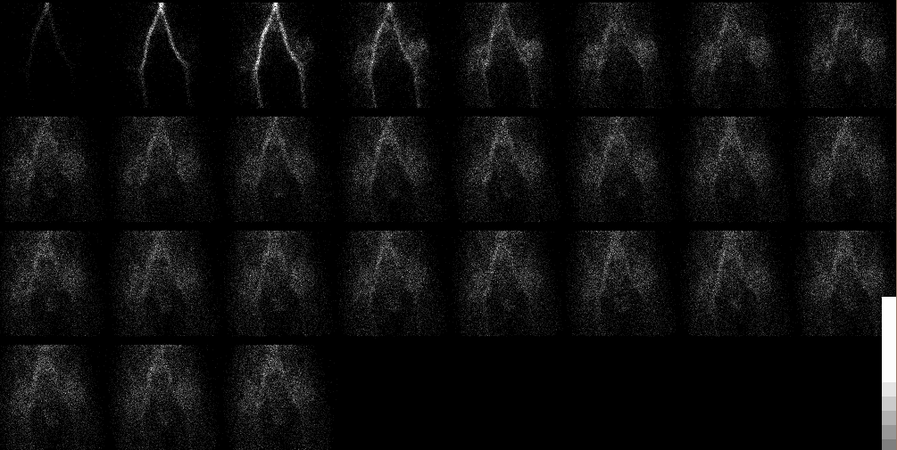
Anterior Radionuclide Angiographic Images of the Pelvis
View main image(bs) in a separate image viewer
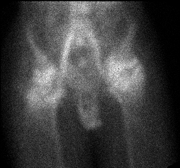
View second image(bs). Anterior Immediate Static Image of the Pelvis
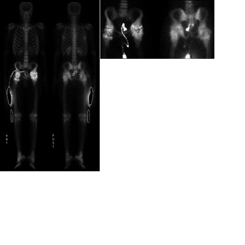
View third image(bs). Delayed Anterior and Posterior Whole Body and Pelvic Spot Images
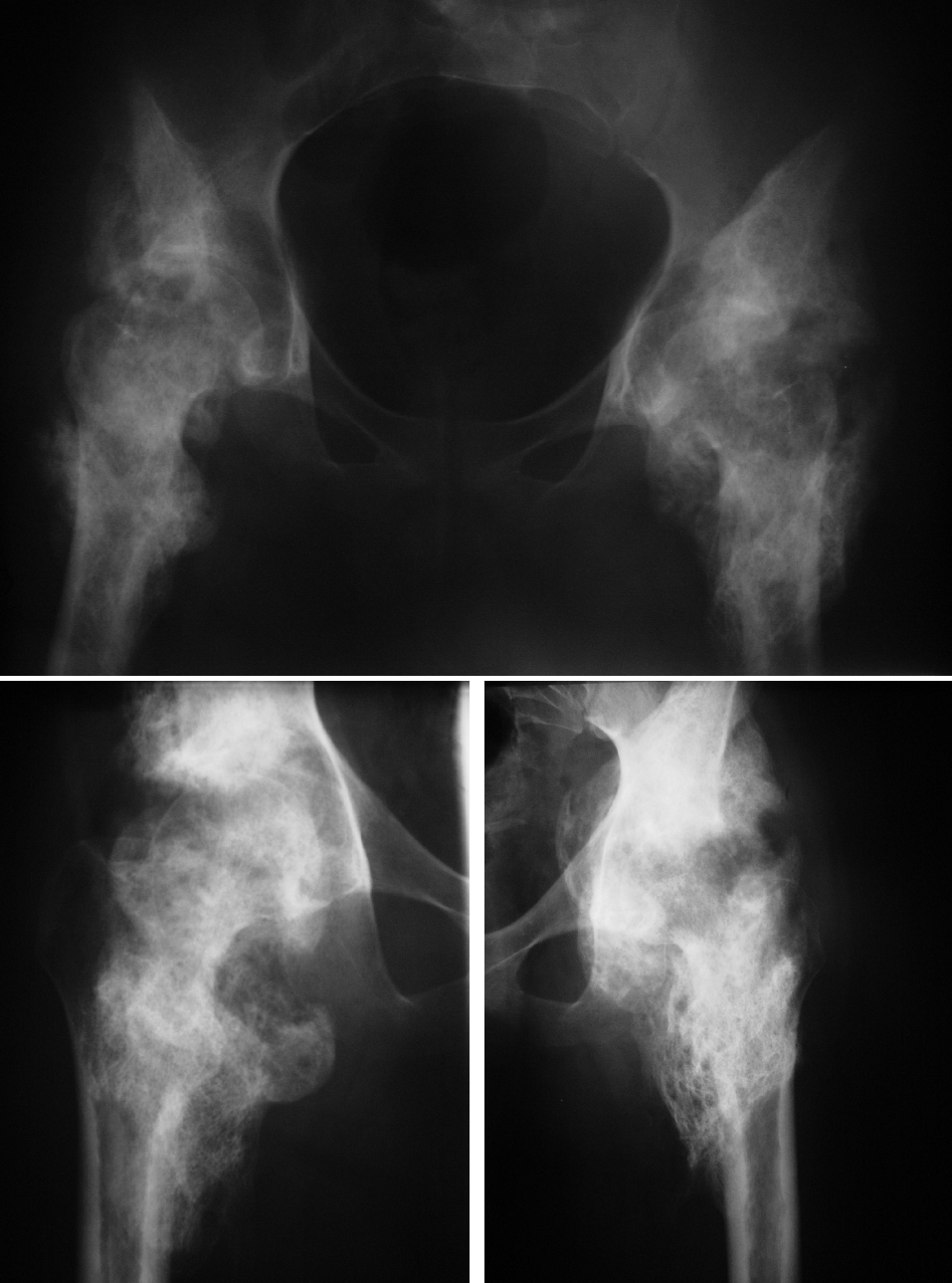
View fourth image(xr). Plain Radiographs of the Pelvis and Hips
Full history/Diagnosis is available below
Radiographic evalution initially demonstrates ill-defined periarticular radiodensities which enlarge and merge with underlying bone. Eventually these may develop trabecular architecture and cause bridging across the joint.
Bone scintigraphy is often used to evaluate the maturity of ossification prior to surgical intervention. This evaluation is important because resection of immature heterotopic ossification often results in recurrence. Acutely, increased radiopharmaceutical activity may be seen as early as 2 weeks following injury. During the subacute phase, there is a rapid increase in activity. A chronic active immature phase follows in which there is a steady state of increased activity. Finally, there is a chronic mature phase in which the activity decreases and returns to normal. Surgery should not be performed until uptake begins to diminish.
In this particular patient, there is markedly increase uptake of radiopharmaceutical, and thus, surgical intervention should be postponed. The clinical, radiographic and scintigraphic features in this case are characteristic and no other entitities were considered.
Reference:
Resnick D. Neuromuscular Disorders. In: Resnick D, Niwayama G, eds. Diagnosis of Bone and Joint Disorders, 2nd Ed. Philadelphia: WB Saunders, 1988.
References and General Discussion of Bone Scintigraphy (Anatomic field:Skeletal System, Category:Misc)
Return to the Teaching File home page.