Case Author(s): Bart Rydzewski M.D., Ph.D. and Jerold Wallis M.D. , 8/23/00 . Rating: #D2, #Q3
Diagnosis: Legg-Calve-Perthes disease
Brief history:
4 year-old boy with right lower extremity pain and limping.
Images:
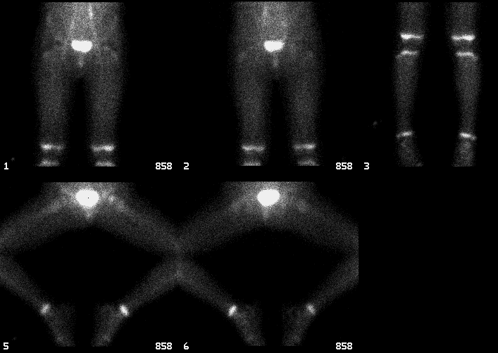
Anterior and frog leg immediate static images of the lower extremities.
View main image(bs) in a separate image viewer
View second image(bs).
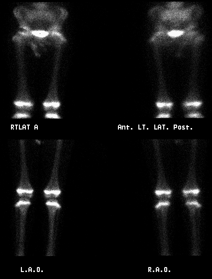
Anterior delayed static images of the lower extremities.
View third image(bs).
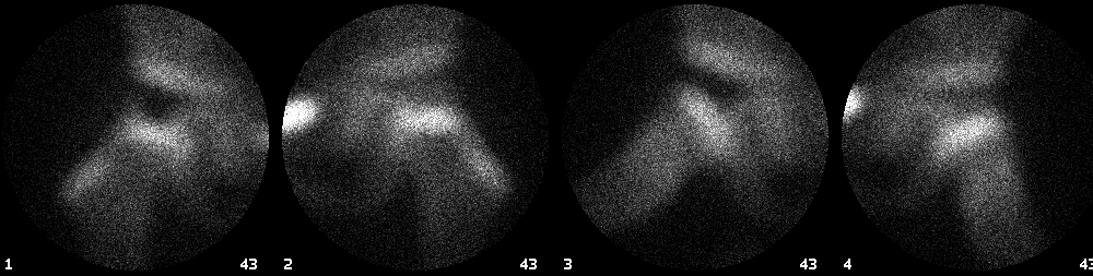
Anterior delayed pinhole images of the hips.
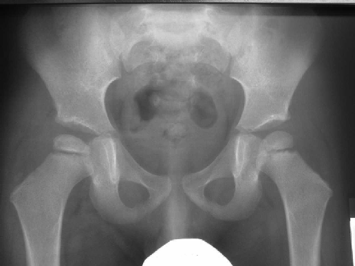
View fourth image(xr).
AP view of the pelvis.
Full history/Diagnosis is available below
Diagnosis: Legg-Calve-Perthes disease
Full history:
4-year-old boy with a history of two weeks of non-specific right lower extremity pain with limping. The pain appear worst with use through the day. The patient has been afebrile and otherwise appears well.
Ultrasonographic examination demonstrated no right hip joint effusion.
Radiopharmaceutical:
5.8 mCi Tc-99m MDP i.v.
Findings:
A limited examination of the lower extremities was performed consisting of immediate and delayed views of the lower extremities and pinhole AP and frog leg views of the hips.
Immediate static images of the hips suggest decreased activity in the region of the right femoral head. Delayed images confirm an area of markedly decreased uptake in the lateral right capital femoral epiphysis and no other abnormalities. This finding is most consistent with Legg-Calve-Perthes disease of the right femoral head.
A plain radiograph of the pelvis demonstrate slightly sclerotic right femoral capital epiphysis which is smaller than the left one. This finding is consistent with the bone scintigraphy study interpretation.
Discussion:
Decreased Tc-99m MDP activity in a femoral head in a pediatric patient may be related to the presence of a hip effusion (with increased joint pressure causing decreased regional blood flow) or Legg-Calve-Perthes (LCP) disease. Hip joint effusion may in turn be related to toxic synovitis or septic arthritis. Since the ultrasonographic examination of the hip in this case did not demonstrate any joint effusion, both of these conditions were rendered very unlikely thus strongly suggesting LCP.
Legg-Calve-Perthes disease is a form of avascular necrosis of the femoral head occurring mostly in children 5 to 10 years old. The condition is generally regarded as idiopathic and it may be related to prior episodes of toxic synovitis. In the early stages of the disease plain radiographs may be normal. At the same time, delayed bone scintigraphic images demonstrate complete absence of activity within the proximal femoral epiphysis. In the later stages of the disease partial revascularization of the head occurs. Classically, it is associated with initial return of activity to the lateral aspect of the femoral head – it is so called “lateral column” sign. Further healing is demonstrated by extension of activity within the center of the femoral head.
Although MRI may currently be the most widely used technique to evaluate LCP disease, bone scintigraphy may also be used, as demonstrated in this case.
References:
Clinical Nuclear Medicine Ed. MN Maisey, KE Britton, BD Collier Chapman and Hall Medical 3rd edition 1998
Textbook of Nuclear Medicine ed. Michael A. Wilson, Lippincott-Raven Publishers 1998
ACR Codes and Keywords:
References and General Discussion of Bone Scintigraphy (Anatomic field:Skeletal System, Category:Metabolic, endocrine, toxic)
Search for similar cases.
Edit this case
Add comments about this case
Return to the Teaching File home page.
Case number: bs120
Copyright by Wash U MO