Case Author(s): Daniel E. Appelbaum, MD and Henry D. Royal, MD , 5/10/00 . Rating: #D2, #Q3
Diagnosis: Multiple thoracic fractures.
Brief history:
30 year old female status post motor vehicle collision four weeks ago.
Images:

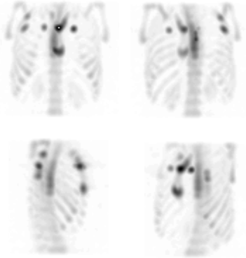
Anterior, posterior, and selected spot images are shown. What are the findings? What else could be performed?
View main image(bs) in a separate image viewer
View second image(bs).
Selected 3D reprojection images from the SPECT reconstruction.
Full history/Diagnosis is available below
Diagnosis: Multiple thoracic fractures.
Full history:
This is a 30 year old female, four weeks status post MVA, who presented to the nuclear medicine department with continued pain in the sternum, back, and right ribs. Plain films acquired over the previous month demonstrated a segond fracture of the left knee and a minimal anterior wedge deformity of the T10 vertebral body. However, there was no radiographic correlate for the patient's continued thoracic skeletal pain. Bone scintigraphy was requested for further evaluation.
Radiopharmaceutical:
20.8 mCi Tc-99m MDP i.v.
Findings:
The whole body and spot planar images demonstrate symmetric, increased radiotracer activity involving both anterior second ribs, near the costochondral junctions. Abnormal activity is also present at the posterolateral right 4th and 6th ribs, the sternal-manuibrial junction, the sternal-xiphoid junction, and two foci of increased activity at the midline of the mid-thoracic spine, possibly localized within the spinous processes. The sternum obscures the thoracic spine on the anterior view, and the oblique spot images do not add anything further. (Note also the abnormal activity at the proximal left tibia laterally as well as at the lateral tibial plateau on the right. There is vague soft tissue activity in the left thigh and calf, also related to trauma suffered in the MVA).
The SPECT images confirm the spinous process localization at T5 and T7. In addition, however, they demonstrate diffuse increased activity involving the T5-T10 vertebral bodies anteriorly which was not at all evident on the planar images.
Discussion:
SPECT imaging during bone scintigraphy can dramatically increase lesion conspicuity and improve localization.
ACR Codes and Keywords:
References and General Discussion of Bone Scintigraphy (Anatomic field:Skeletal System, Category:Effect of Trauma)
Search for similar cases.
Edit this case
Add comments about this case
Return to the Teaching File home page.
Case number: bs116
Copyright by Wash U MO