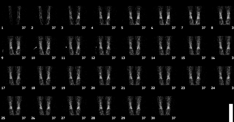

Anterior tibial/ankle flow images
View main image(bs) in a separate image viewer
View second image(bs). Anterior tibial/ankle immediate static images
View third image(bs). Anterior knee/tibial/ankle immediate static images
View fourth image(bs). Delayed whole-body images
Full history/Diagnosis is available below
There is also increased uptake in the majority of the ulnae radii bilaterally, in the posterior left rib, in the anterior aspect of the first right rib, and in the proximal aspect of the left humerus.
Plain radiographs of these areas demonstrate corresponding abnormalities consisting of scalloping of the cortex and a cystic and sclerotic mixed medullary pattern with thickened trabeculae.
Age: 50-70y
Clinical: Ranges from joint pain --> systemic involvement (heart, liver, spleen, pancreas, pericardium, lungs, adrenals, aorta, lymph nodes, bowel, orbit, and bone)
Histology: Replacement of bone marrow by foamy lipid, histiocytes, and giant cells causing medullary fibrosis and osteosclerosis
X-ray: Symmetric, appendicular skeleton >> axial skeleton Long bones invariably affected in the diaphysis and metaphysis with:
- Patchy or diffuse increase in trabecular pattern
- Medullary sclerosis
- Cortical thickening
Nucs: Increased uptake with Tc-99m MDP and Gallium-67. Photopenia with In-111 and Tc-99m SC
Prognosis: 1/3 Fatal
Treatment: Radiation, chemotherapy, steroids
View followup image(xr). There are multiple small mixed, lytic, and sclerotic lesions throughout the tibia, primarily located in the ends of the diaphysis and the metaphysis of the tibia. There is also a small lytic lesion in the proximal fibular diaphysis, as well as in the distal femur. When compared to the previous plain xrays examinations, there has been no significant interval change. These areas of abnormality correlate with the areas of increased radiopharmaceutical uptake on the bone scan.
References and General Discussion of Bone Scintigraphy (Anatomic field:Skeletal System, Category:Other generalized systemic disorder)
Return to the Teaching File home page.