Case Author(s): David A. Hillier, M.D., Ph.D. and Tom R. Miller, M.D., Ph.D. , 6/99 . Rating: #D2, #Q4
Diagnosis: Stroke on bone scintigraphy
Brief history:
57 year-old man with a right upper lobe mass.
Images:
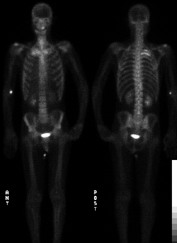
Bone scintigraphy
View main image(bs) in a separate image viewer
View second image(ct).
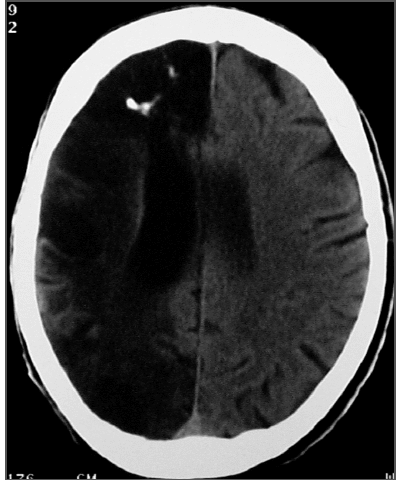
Head computed tomography
View third image(bs).
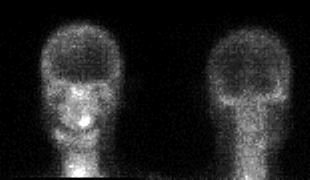
Enlarged spot images of the head.
Full history/Diagnosis is available below
Diagnosis: Stroke on bone scintigraphy
Full history:
57 year-old man with a right upper lobe mass. A right neck mass was resected in 1997, complicated by right carotid and internal jugular hemorrhage and subsequent ligation. Postoperatively the patient developed left hemiparesis. Right hemispheric edema, consistent with infarct, was seen on CT.
Radiopharmaceutical:
21.0 mCi Tc-99m MDP, i.v.
Findings:
Bone scintigraphy:
- Intense uptake in posterior right 3rd and 4th ribs.
- Linear uptake along the lateral and medial margins of the femora and tibiae.
- Mildly increased uptake overlying right cerebrum, likely related to the patient’s H/O right CVA.
(2. CT of the chest (not shown))
- 3.5 x 2 cm soft tissue mass abutting pleura in the right lung apex.
3. Computed tomography of the head:
- Extensive right cerebral encephalomalacia with foci of calcification, consistent with old infarct.
Discussion:
Although uptake of a bone imaging agent is not commonly seen in patients with stroke, in this case, there is severe right hemispheric encephalomalacia with calcification on CT.
ACR Codes and Keywords:
References and General Discussion of Bone Scintigraphy (Anatomic field:Skeletal System, Category:Misc)
Search for similar cases.
Edit this case
Add comments about this case
Return to the Teaching File home page.
Case number: bs114
Copyright by Wash U MO