Case Author(s): Lester Johnson, M.D., Ph.D. and Jerold Wallis, M.D. , 07/26/99 . Rating: #D2, #Q4
Diagnosis: Soft tissue uptake after radiation therapy
Brief history:
Evaluation for progression of metastatic breast cancer
Images:

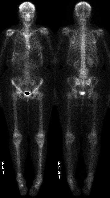
Anterior and posterior whole body images
View main image(bs) in a separate image viewer
View second image(bs).
Anterior and posterior whole body images obtained 6 months earlier.
The posterior image is repeated at a brighter intensity.
Full history/Diagnosis is available below
Diagnosis: Soft tissue uptake after radiation therapy
Full history:
This 40 year old woman has a history of known metastatic breast carcinoma. A bone scan obtained nine months earlier demonstrated several skeletal metastases including increased uptake in L4 (not shown). At that time she complained of increasing low back pain and was treated with external beam radiation therapy (XRT) to her lumbar spine. Three months after her XRT, she received the bone scan shown second above. Nine months after the XRT she received the bone scan shown first above (the unknown).
Radiopharmaceutical:
99m-Technetium-HDP
Findings:
Nine months after her XRT, the patient received the bone scan shown first above which demonstrates abnormally increased accumulation of radiotracer in a rectangular configuration around the lumbar spine (several osseous metastases are also again noted). This pattern of uptake around the lumbar spine is clearly not osseous but, rather, represents soft tissue uptake. The location of soft tissue uptake matches the XRT port shown below.
Discussion:
The diagnosis of post-XRT soft tissue uptake should be suggested based on the unusual geometric configuration (rectangle) of the uptake--it is difficult to imagine how else to explain this pattern which otherwise makes no anatomic sense. The presence of osseous metastatic disease in the study further supports the supposition that the patient might have received XRT (in the absence of knowing this history).
View followup image(xr).
Lumbar spine radiograph demonstrates radiation therapy port
Major teaching point(s):
Soft tissue uptake of diphosphonate radiotracers is not unusual and is often associated with states of abnormal calcium deposition in soft tissues (e.g., heterotopic bone formation, dystrophic soft tissue calcification). Increased soft tissue accumulation of bone scanning agents is also associated with tissue infarction and necrosis (e.g., myocardial infarction, which is the basis for pyrophosphate scanning to diagnose acute/subacte myocardial infarction, and cerebral infarction). The mechanism of uptake in infarction may be influx of extracellular calcium into dying cells. The mechanism of soft tissue uptake after XRT is not known, but has been postulated to be ischemic necrosis induced by the XRT. What makes this case unusual is the intensity of the soft tissue uptake so late after the XRT (nine months) together with the way in which the soft tissue uptake increased between 3 months and 9 months after XRT.
ACR Codes and Keywords:
- General ACR code: 48
- Skeletal System:
4.81 "CALCIFICATION, OSSIFICATION"
References and General Discussion of Bone Scintigraphy (Anatomic field:Skeletal System, Category:Misc)
Search for similar cases.
Edit this case
Add comments about this case
Return to the Teaching File home page.
Case number: bs113
Copyright by Wash U MO

