After viewing the image(s), the Full history/Diagnosis is available by using the link here or at the bottom of this page
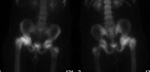
Delayed limited bone scan of the hips and pelvis are shown
View main image(bs) in a separate viewing box
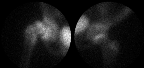
View second image(bs). Pinhole views of the hips are shown.
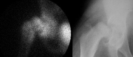
View third image(xr). A right hip radiograph is shown.
View fourth image(mm). Side-by-side view of the right hip on bone scintigram and plain radiograph
Full history/Diagnosis is also available
Return to the Teaching File home page.