Case Author(s): David A. Hillier, M.D., Ph.D. and Henry Royal, M.D. , . Rating: #D4, #Q4
Diagnosis: Engelmann's disease (progressive diaphyseal dysplasia)
Brief history:
Thirty-eight-year-old woman with a known history of a skeletal dysplasia.
Images:
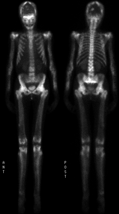
Bone scintigraphy
View main image(bs) in a separate image viewer
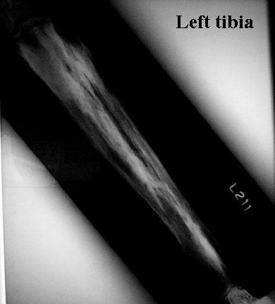
View second image(xr).
Plain film of tibia
Full history/Diagnosis is available below
Diagnosis: Engelmann's disease (progressive diaphyseal dysplasia)
Full history:
Thirty-eight-year-old woman with a known history of a skeletal dysplasia.
Radiopharmaceutical:
Bone scintigraphy, 21.1 mCi methylene diphosphonate
Findings:
1. Bone scintigraphy:
- Diffusely increased uptake throughout the skeleton. Hands and feet are relatively spared.
- Focal intense foci in skull, proximal humeri, femora and tibiae.
2. Skull, femur plain films:
- Sclerotic undertubulated bones.
- Sclerotic changes throughout the skull, especially the skull base.
- Lytic areas corresponding to foci of intense uptake on bone scan.
Discussion:
Progressive diaphyseal dysplasia (Engelmann’s disease) is hereditary. The long bones become undertubulized and the medullary cavity becomes narrowed.
Differential Diagnosis List
This appearance, with diffusely increased uptake throughout the skeleton with undertubulation of the bones is characteristic of Engelmann’s disease.
ACR Codes and Keywords:
References and General Discussion of Bone Scintigraphy (Anatomic field:Skeletal System, Category:Misc)
Search for similar cases.
Edit this case
Add comments about this case
Read comments about this case
Return to the Teaching File home page.
Case number: bs109
Copyright by Wash U MO