Case Author(s): David A. Hillier, M.D., Ph.D. and Mark Mintun, M.D. , 7/3/99 . Rating: #D2, #Q3
Diagnosis: delete - Cellulitis
Brief history:
48 year-old man with ulceration of dorsal mid right foot.
Images:
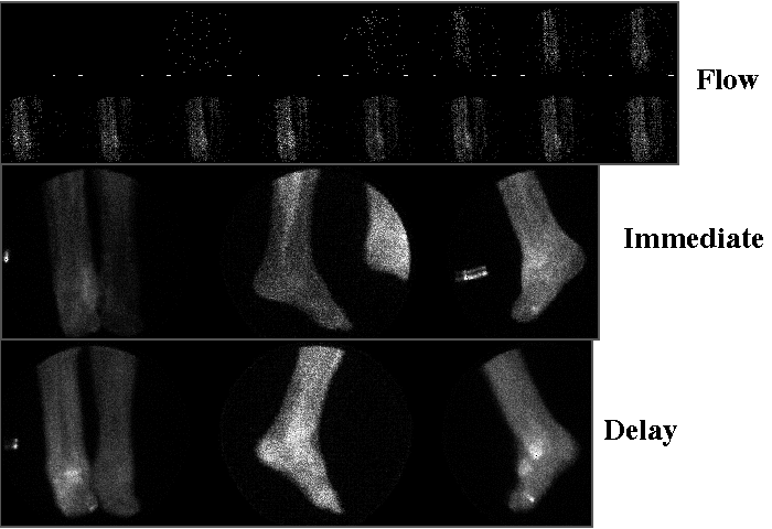
Three phase bone scintigraphy of feet
View main image(bs) in a separate image viewer
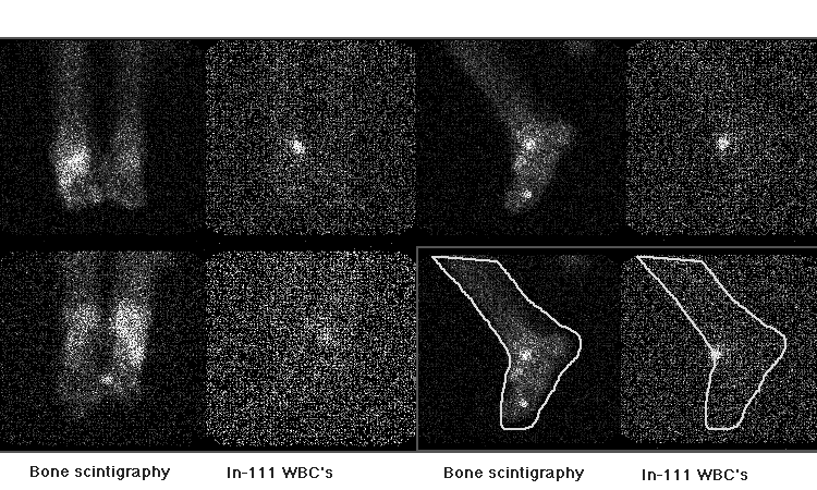
View second image(iw).
Bone scintigraphy and In-111 WBC
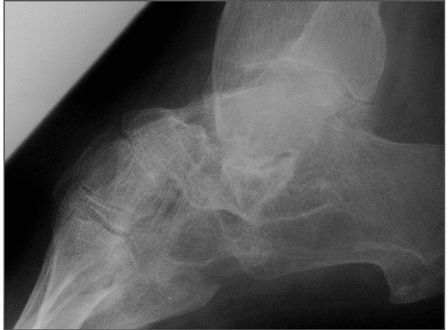
View third image(xr).
Lateral right foot plain film
Full history/Diagnosis is available below
Diagnosis: delete - Cellulitis
Full history:
48 year-old man with ulceration of dorsal mid right foot.
Radiopharmaceutical:
Bone scintigraphy and Leukocyte scintigraphy, 20.5 mCi Tc-99m MDP and 433 mCi In-111 labeled autologous leukocytes, IV
Findings:
1. Bone scintigraphy and leukocyte scintigraphy:
- In-111 WBC uptake anterior to right tibiotalar joint consistent with infection.
- No evidence of osteomyelitis.
- Increased uptake in right talus, corresponding to collapse on plain film.
- Increased uptake on bone scintigraphy in right 1st toe, consistent with degenerative / post surgical changes.
2. Ankle and foot plain films:
- Bilateral club foot and hammer toe. Diffuse osteopenia. Collapse of right talar dome (appears chronic).
Discussion:
In-111 WBC or gallium imaging may be added to standard bone scintigraphy is complex cases to improve diagnostic accuracy. In gallium imaging, comparison is made with the bone scintigraphy to judge the relative uptake of gallium with respect to bone. If one calls osteomyelitis when the two reveal equal degrees of uptake, sensitivity is good, but specificity suffers. If one requires that the gallium uptake be higher, then sensitivity is reduced at the expense of improved specificity.
Any focus of increased In-111 WBC should be considered suspicious.
Differential Diagnosis List
On bone scintigraphy there are several foci of increased uptake. It is difficult, in this patient with congenital foot deformities and degenerative changes to rule out osteomyelitis. Therefore In-111 WBC imaging was added. Careful analysis of the images reveals a single focus of increased uptake on leukocyte imaging which is anterior the bones, consistent with cullilitis
ACR Codes and Keywords:
References and General Discussion of Bone Scintigraphy (Anatomic field:Skeletal System, Category:Inflammation,Infection)
Search for similar cases.
Edit this case
Add comments about this case
Return to the Teaching File home page.
Case number: bs107
Copyright by Wash U MO