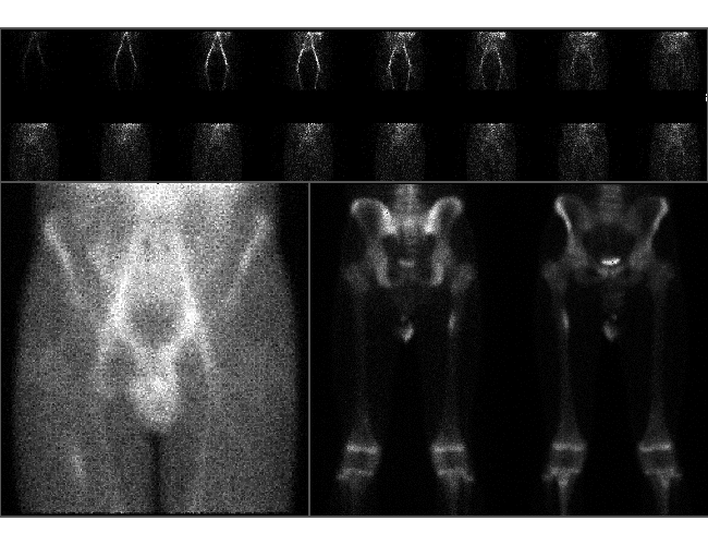

Bone scintigraphy
View main image(bs) in a separate image viewer
View second image(xr). Right femur plain films
Full history/Diagnosis is available below
- Ovoid focal uptake at medial aspect of proximal right femoral metadiaphysis.
2. Right femur:
- Periosteal formation correlating with scintigraphic abnormality.
Bone scintigraphy becomes positive at the same time or sooner than plain films and has a low false-negative rate. The most common sites of stress fracture are the femoral neck and tibia. Less commonly, the femoral diaphysis may be involved.
References:
Burr D, et al. Experimental stress fractures of the tibia. J of Bone & Joint Surg. 72-B, 370-375. 1990.
Donohoe K. Selected topics in orthopedic nuclear medicine. Orthop clin N Am. 29, 85-101. 1998.
Holder L. Bone scintigraphy in skeletal trauma. Rad clin N Am. 31, 739-781. 1993.
Lesho E. Can tuning forks replace bone scans for identification of tibial stress fractures? Milit Med. 162, 802-803.
Milgrom C, et al. The long-term followup of soldiers with stress fractures. 13, 398-400. 1985.
Wilcox J, et al. Bone scanning in the evaluation of exercise-related stress injuries. Rad. 123, 699-703. 1977.
Zwas ST, et al. Interpretation and classification of bone scintigraphic findings in stress fractures. J Nucl Med. 28, 452-457. 1987.
References and General Discussion of Bone Scintigraphy (Anatomic field:Skeletal System, Category:Effect of Trauma)
Return to the Teaching File home page.