Case Author(s): David A. Hillier, M.D., Ph.D. and Farroqh Dehdashti, M.D. , 6/28/99 . Rating: #D2, #Q4
Diagnosis: Hypertrophic osteoarthropathy
Brief history:
76 year-old male with history of non-small-cell lung carcinoma.
Images:

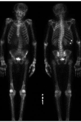
Bone scintigraphy, anterior and posterior images.
View main image(bs) in a separate image viewer
View second image(ct).
Computed tomography
View third image(bs).
Bone scintigraphy before and after therapy
Full history/Diagnosis is available below
Diagnosis: Hypertrophic osteoarthropathy
Full history:
77 yo man with history of a right lobectomy for lung non-small cell lung carcinoma who now presents with a new large left upper lobe mass. He subsequently underwent radiation therapy to the chest wall.
Radiopharmaceutical:
20.6 mCi MDP
Findings:
1. Bone scintigraphy:
- Posterior left 4th and 5th rib uptake.
- Increased uptake in the distal radius, ulna hands and periosteal uptake in the tibiae.
- Foci of uptake, consistent with rib fractures of the right 10th and 11th ribs.
2. Computed tomography of the chest:
- 6x5x5 cm mass in the left upper lobe with necrotic center.
- Right lung scar, consistent with H/O right upper lobectomy.
3. Bone scintigraphy 9 months later:
- Decreased left 4th and 5th rib uptake, consistent with H/O interval radiation.
- Essentially resolved hypertrophic osteoarthropathy.
4. Computed tomography of the chest 9 months later:
- Diffuse fibrotic and volume loss changes in the left lung consistent with XRT.
- Lenticular low attenuation at site of prior tumor (necrotic tumor vs loculated effusion).
- New necrotic lesion at base of left neck worrisome for metastatic disease.
Discussion:
Hypertrophic osteoarthropathy (HOA) was initially described by Bamberger in 1889 and then by Marie in 1890. It is still known as Bamberger-Marie syndrome in europe. The primary form, primary familial hypertrophic osteoarthropathy (pachydermoperiostitis) is an autosomal dominant trait. The secondary form can result from a number of extraskeletal pathologies, particularly pulmonary (hence the former term for this syndrome, pulmonary hypertrophic osteoarthropathy). Some consider hypertrophic osteoarthropathy and pachydermoperiostis as seperate entities and that HOA is, by definition, due to secondary causes. Patients typically present with tenderness in the long bones. There is periosteal proliferation and formation of new bone. Bone remodelling at the distal phalangeal tufts results in clubbing, which is nearly always seen with hypertrophic osteoarthropathy. Bone scintigraphy is more sensitive than plain film in detection of the new bone formation.
The etiology of hypertrophic osteoarthropathy is poorly understood. There are several theories, including:
1) Neuronal. Afferent signal via vagus or intercostal nerves signal an efferent nerve or hormonal response.
2) Hormonal. Some tumors produce a substance that results in periosteal reaction.
3) A-V shunt. Lung normally metabolizes substances that can cause periosteal reaction. If there is lung disease, a shunt can develop that allows these substances through.
Differential Diagnosis List
The differential diagnosis of periosteal bone scintigraphy uptake includes hypertrophic osteoarthropathy, metabolic causes (prostaglandin therapy, scurvy, healing rickets, hypervitaminosis A and D, thyroid acropachy), peripheral vascular disease and pachydermoperiostitis. In this case, there is evidence of a lung cancer on bone scintigraphy and hypertrophic osteoarthropathy is likely. The subsequent time course with near-resolution of the findings with treatment of the tumor verifies the etiology.
ACR Codes and Keywords:
- General ACR code: 48
- Skeletal System:
4.861 "Hypertrophic osteoarthropathy"
References and General Discussion of Bone Scintigraphy (Anatomic field:Skeletal System, Category:Misc)
Search for similar cases.
Edit this case
Add comments about this case
Read comments about this case
Return to the Teaching File home page.
Case number: bs105
Copyright by Wash U MO

