
Bone scintigraphy
View main image(bs) in a separate image viewer
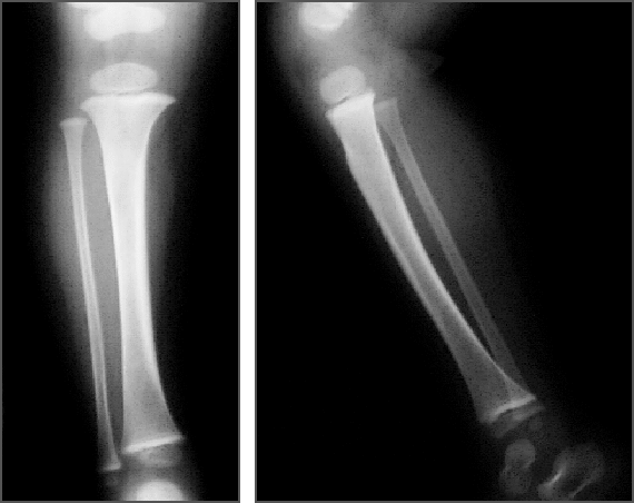
View second image(xr). Radiographs of right tibia
Full history/Diagnosis is available below
- Anterior radionuclide angiogram and immediate static image reveal mild hyperperfusion and somewhat greater hyperemia in the region of the entire right tibial shaft.
- Anterior and right and left frog-leg lateral delayed images show moderately increased uptake in the right tibia (except the proximal metaphyseal region)
2. Right leg radiographs (five days earlier):
- Normal
If radiographs in a case of suspected toddler’s fracture are negative, a cast will commonly be placed for 7 to 10 days. This is then removed and a new radiograph is obtained. This will usually reveal the fracture or periosteal reaction if there has been a spiral fracture.
If there is a concomitant fever, leukocytosis or other reason to suspect an inflammatory process, bone scintigraphy should be considered. The uptake pattern in toddler's fracture will typically reveal a linear, spiral band of increased activity or more diffuse uptake that is centered in the mid diaphysis, as in this case (Miller and Sanderson, 1998).
Bonfield W, Grynpas M. Spiral fracture of cortical bone. Biomechanics. 15, 555-559. 1982.
Chao S, et al. A mechanism of spiral fracture of the humerus: a report of 129 cases following the throwing of hand grenades. J Trauma. 7, 602-605. 1971.
Hieilbronner D, et al. Fractures of the humerus in arm wrestlers. Clin Orthop and Related Res.149, 169-171. 1980.
Miller J, Sanderson R. Scintigraphy of toddler’s fracture. J Nucl Med. 29, 2001-2003. 1998.
References and General Discussion of Bone Scintigraphy (Anatomic field:Skeletal System, Category:Effect of Trauma)
Return to the Teaching File home page.