Case Author(s): John R. Leahy, M.D. and Tom R. Miller, M.D., Ph.D. , 6/20/99 . Rating: #D2, #Q3
Diagnosis: Hypertrophic osteoarthropathy and Lung carcinoma metastasis
Brief history:
Lung cancer evaluation for metastases.
Images:

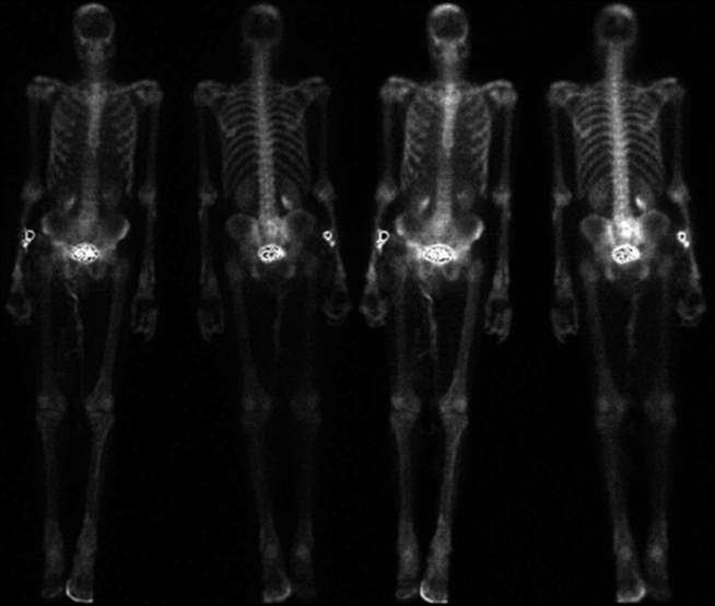
Dual intensity whole body delay images.
View main image(bs) in a separate image viewer
View second image(bs).
Spot views of the head and chest, along with anterior views of the feet
after washing.
Full history/Diagnosis is available below
Diagnosis: Hypertrophic osteoarthropathy and Lung carcinoma metastasis
Full history:
64 year-old man with newly diagnosed lung carcinoma. The current
study is part of a staging evaluation to assess for osseous metastases.
Radiopharmaceutical:
20.5 mCi Tc-99m MDP i.v.
Findings:
Delayed whole body scintigrams show lesions with increased activity in the
parietal bones, right greater than left.
Increased activity is seen diffusely in the tibiae, ulnae, wrists and
hands in a cortically-based pattern. The rim of activity along the right anterior foot is absent after
washing, and was felt to represent urine contamination.
Discussion:
The activity in the upper and lower extremities is consistent with
hypertrophic osteoarthropathy. This condition typically produces a characteristic
appearance on scintigraphic studies. Increased activity is seen along
the cortical margins of certain long and flat bones. Specific sites of
involvement include the following: tibia and fibula 95%, femur 88%,
hands and wrists 88%, radius and ulna 85%, feet 81%, scapula 67%,
mandible 42%, clavicle 33%, ribs 2%, and pelvis 2%. Activity is usually
centered in the diaphases, but can be found in a juxta-articular
distribution, especially in the phalanges. Asymmetric involvement occurs
in 15% of patients. The scintigraphic findings often precede radiographic
findings, and may regress with treatment of the primary etiology.
This condition is seen in approximately 5% of lung carcinoma patients,
as well as other pulmonary malignancies such as mesothelioma.
It is also seen in non-malignant pulmonary conditions such as COPD,
abscess or pleural fibroma. Extra pulmonary etiologies include cyanotic
congenital heart disease, inflammatory diseases of the GI tract, and
malabsorption syndromes.
Activity in the parietal bones was felt to represent osseous metastases as well as possible sacral metastasis.
References:
Sandler MP, et al. Diagnostic Nuclear Medicine, 3rd ed 1996. Williams and
Wilkins, Baltimore. p 650-51.
Wagner HN, et al. Principles of Nuclear Medicine 2nd ed 1995. W.B. Saunders
Company, Philadelphia. p 1005-1006
ACR Codes and Keywords:
References and General Discussion of Bone Scintigraphy (Anatomic field:Skeletal System, Category:Neoplasm, Neoplastic-like condition)
Search for similar cases.
Edit this case
Add comments about this case
Return to the Teaching File home page.
Case number: bs103
Copyright by Wash U MO

