Case Author(s): Tate Allen, M.D. and Tom R. Miller, M.D., Ph.D. , 10/9/98 . Rating: #D2, #Q3
Diagnosis: Osteomyelitis
Brief history:
This is a 52 year-old diabetic with a foot ulcer.
Images:
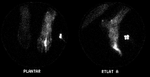
Delayed Bone Scintigraphy
View main image(bs) in a separate image viewer
View second image(bs).
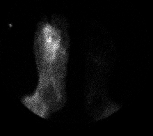
Plantar Flow Images
View third image(bs).
Plantar Immediate Static Image
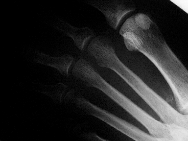
View fourth image(xr).
Right Foot Plain Film
Full history/Diagnosis is available below
Diagnosis: Osteomyelitis
Full history:
This is a 52 year-old diabetic man with a right plantar ulcer over the 3rd digit
which has been present for three months. Recently, the patient
has had a low grade fever and worsening foot pain.
Radiopharmaceutical:
21.3 mCi Tc-99m MDP i.v.
Findings:
There is increased blood flow to the right forefoot with increased
blood pool activity.
Delayed images show marked uptake
at the right third distal metatarsal and proximal phalanx. These
findings are consistant with osteomyelitis of these bones and possible
third metatarsal-phalangeal joint infection. In addition,
there is diffusely increased activity in the right foot soft tissue and bones. This is consistant with hyperemia and
cellulitis. The plain
films show no osseous abnormalities.
Discussion:
Bone scintigraphy is commonly abnormal before plain film findings are
apparent.
Followup:
The patient was started on i.v. antibiotics.
ACR Codes and Keywords:
- General ACR code: 42
- Skeletal System:
4.21 "OSTEOMYELITIS (for 4th number see box following .219)"
References and General Discussion of Bone Scintigraphy (Anatomic field:Skeletal System, Category:Inflammation,Infection)
Search for similar cases.
Edit this case
Add comments about this case
Return to the Teaching File home page.
Case number: bs096
Copyright by Wash U MO